 Movie
Movie Controller

[English] 日本語
 Yorodumi
- PDB-8apv: Crystal Structure of H. influenzae TrmD in complex with Compound 27 -
+ Open data

- Basic information
| Entry | Database: PDB / ID: 8apv | ||||||
|---|---|---|---|---|---|---|---|
| Title | Crystal Structure of H. influenzae TrmD in complex with Compound 27 | ||||||
 Components Components | tRNA (guanine-N(1)-)-methyltransferase TRNA (guanine9-N1)-methyltransferase TRNA (guanine9-N1)-methyltransferase | ||||||
 Keywords Keywords | RNA BINDING PROTEIN / Methyltransferase | ||||||
| Function / homology |  Function and homology informationtRNA (guanine37-N1)-methyltransferase / tRNA (guanine(37)-N1)-methyltransferase activity / tRNA modification / methylation / cytoplasm Function and homology informationtRNA (guanine37-N1)-methyltransferase / tRNA (guanine(37)-N1)-methyltransferase activity / tRNA modification / methylation / cytoplasmSimilarity search - Function | ||||||
| Biological species |  Haemophilus influenzae (bacteria) Haemophilus influenzae (bacteria) | ||||||
| Method | X-RAY DIFFRACTION / MOLECULAR REPLACEMENT / Resolution: 2.4 Å | ||||||
 Authors Authors | Hall, G. / Cowan, R. / Carr, M.D. | ||||||
| Funding support |  United Kingdom, 1items United Kingdom, 1items
| ||||||
 Citation Citation | Journal: Bioorg.Med.Chem.Lett. / Year: 2023 Title: Evaluating the druggability of TrmD, a potential antibacterial target, through design and microbiological profiling of a series of potent TrmD inhibitors. Authors: Wilkinson, A.J. / Ooi, N. / Finlayson, J. / Lee, V.E. / Lyth, D. / Maskew, K.S. / Newman, R. / Orr, D. / Ansell, K. / Birchall, K. / Canning, P. / Coombs, P. / Fusani, L. / McIver, E. / ...Authors: Wilkinson, A.J. / Ooi, N. / Finlayson, J. / Lee, V.E. / Lyth, D. / Maskew, K.S. / Newman, R. / Orr, D. / Ansell, K. / Birchall, K. / Canning, P. / Coombs, P. / Fusani, L. / McIver, E. / Pisco, J. / Ireland, P.M. / Jenkins, C. / Norville, I.H. / Southern, S.J. / Cowan, R. / Hall, G. / Kettleborough, C. / Savage, V.J. / Cooper, I.R. | ||||||
| History |
|
- Structure visualization
| Structure viewer | Molecule: MolmilJmol/JSmol |
|---|
- Downloads & links
-Download
| PDBx/mmCIF format | 8apv.cif.gz | 113.8 KB | Display | PDBx/mmCIF format |
|---|---|---|---|---|
| PDB format | pdb8apv.ent.gz | 85.6 KB | Display | PDB format |
| PDBx/mmJSON format | 8apv.json.gz | Tree view | PDBx/mmJSON format | |
| Others |  Other downloads Other downloads |
-Validation report
| Arichive directory | https://data.pdbj.org/pub/pdb/validation_reports/ap/8apvftp://data.pdbj.org/pub/pdb/validation_reports/ap/8apv | HTTPS FTP |
|---|
-Related structure data
| Related structure data |  8aptC 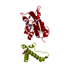 8apuC  8apwC  4yvhS S: Starting model for refinement C: citing same article ( |
|---|---|
| Similar structure data |
-Links
 PDBj
PDBj- Assembly
| Deposited unit | 
| ||||||||
|---|---|---|---|---|---|---|---|---|---|
| 1 | 
| ||||||||
| Unit cell |
|
-Components
| #1: Protein | TRNA (guanine9-N1)-methyltransferase / M1G-methyltransferase / tRNA [GM37] methyltransferase Mass: 29750.016 Da / Num. of mol.: 1 Source method: isolated from a genetically manipulated source Source: (gene. exp.) Haemophilus influenzae (bacteria) / Strain: PittGG / Gene: trmD, CGSHiGG_03625 / Production host: Escherichia coli (E. coli)References: UniProt: A5UG04, tRNA (guanine37-N1)-methyltransferase |
|---|---|
| #2: Chemical | ChemComp-NLL /   Mass: 280.324 Da / Num. of mol.: 1 / Source method: obtained synthetically / Formula: C16H16N4O / Feature type: SUBJECT OF INVESTIGATION Mass: 280.324 Da / Num. of mol.: 1 / Source method: obtained synthetically / Formula: C16H16N4O / Feature type: SUBJECT OF INVESTIGATION |
| #3: Chemical | ChemComp-CIT / Citric acid  Mass: 192.124 Da / Num. of mol.: 1 / Source method: obtained synthetically / Formula: C6H8O7 Mass: 192.124 Da / Num. of mol.: 1 / Source method: obtained synthetically / Formula: C6H8O7 |
| #4: Water | ChemComp-HOH / Water Mass: 18.015 Da / Num. of mol.: 94 / Source method: isolated from a natural source / Formula: H2O Mass: 18.015 Da / Num. of mol.: 94 / Source method: isolated from a natural source / Formula: H2O |
| Has ligand of interest | Y |
-Experimental details
-Experiment
| Experiment | Method: X-RAY DIFFRACTION / Number of used crystals: 1 |
|---|
- Sample preparation
| Crystal | Density Matthews: 2.64 Å3/Da / Density % sol: 53.48 % |
|---|---|
| Crystal grow | Temperature: 291 K / Method: vapor diffusion, hanging drop / pH: 7.5 Details: 20% PEG3350 0.1 M HEPES, pH 7.5 0.1 M potassium citrate tribasic |
-Data collection
| Diffraction | Mean temperature: 100 K / Serial crystal experiment: N |
|---|---|
| Diffraction source | Source: ROTATING ANODE / Type: RIGAKU MICROMAX-007 HF / Wavelength: 1.5418 Å |
| Detector | Type: RIGAKU SATURN 944+ / Detector: CCD / Date: Sep 10, 2020 |
| Radiation | Protocol: SINGLE WAVELENGTH / Monochromatic (M) / Laue (L): M / Scattering type: x-ray |
| Radiation wavelength | Wavelength: 1.5418 Å / Relative weight: 1 |
| Reflection | Resolution: 2.4→47.36 Å / Num. obs: 12287 / % possible obs: 100 % / Redundancy: 6.9 % / CC1/2: 0.994 / Rmerge(I) obs: 0.17 / Rpim(I) all: 0.102 / Rrim(I) all: 0.199 / Net I/σ(I): 11.6 |
| Reflection shell | Resolution: 2.4→2.49 Å / Redundancy: 5.7 % / Rmerge(I) obs: 0.929 / Num. unique obs: 1275 / CC1/2: 0.696 / Rpim(I) all: 0.624 / Rrim(I) all: 1.124 |
- Processing
| Software |
| ||||||||||||||||||||||||||||||||||||||||||||||||||||||||||||||||||||||||||||||||||||||||||||||||||||||||||||||||||||||||||||||||||||||||||||||||||||||
|---|---|---|---|---|---|---|---|---|---|---|---|---|---|---|---|---|---|---|---|---|---|---|---|---|---|---|---|---|---|---|---|---|---|---|---|---|---|---|---|---|---|---|---|---|---|---|---|---|---|---|---|---|---|---|---|---|---|---|---|---|---|---|---|---|---|---|---|---|---|---|---|---|---|---|---|---|---|---|---|---|---|---|---|---|---|---|---|---|---|---|---|---|---|---|---|---|---|---|---|---|---|---|---|---|---|---|---|---|---|---|---|---|---|---|---|---|---|---|---|---|---|---|---|---|---|---|---|---|---|---|---|---|---|---|---|---|---|---|---|---|---|---|---|---|---|---|---|---|---|---|---|
| Refinement | Method to determine structure: MOLECULAR REPLACEMENT Starting model: 4YVH Resolution: 2.4→47.36 Å / Cor.coef. Fo:Fc: 0.952 / Cor.coef. Fo:Fc free: 0.896 / SU B: 7.446 / SU ML: 0.17 / Cross valid method: FREE R-VALUE / ESU R: 0.34 / ESU R Free: 0.26 Details: Hydrogens have been added in their riding positions
| ||||||||||||||||||||||||||||||||||||||||||||||||||||||||||||||||||||||||||||||||||||||||||||||||||||||||||||||||||||||||||||||||||||||||||||||||||||||
| Solvent computation | Ion probe radii: 0.8 Å / Shrinkage radii: 0.8 Å / VDW probe radii: 1.2 Å / Solvent model: MASK BULK SOLVENT | ||||||||||||||||||||||||||||||||||||||||||||||||||||||||||||||||||||||||||||||||||||||||||||||||||||||||||||||||||||||||||||||||||||||||||||||||||||||
| Displacement parameters | Biso mean: 29.48 Å2
| ||||||||||||||||||||||||||||||||||||||||||||||||||||||||||||||||||||||||||||||||||||||||||||||||||||||||||||||||||||||||||||||||||||||||||||||||||||||
| Refinement step | Cycle: LAST / Resolution: 2.4→47.36 Å
| ||||||||||||||||||||||||||||||||||||||||||||||||||||||||||||||||||||||||||||||||||||||||||||||||||||||||||||||||||||||||||||||||||||||||||||||||||||||
| Refine LS restraints |
| ||||||||||||||||||||||||||||||||||||||||||||||||||||||||||||||||||||||||||||||||||||||||||||||||||||||||||||||||||||||||||||||||||||||||||||||||||||||
| LS refinement shell |
|
