 Movie
Movie Controller

[English] 日本語
 Yorodumi
- PDB-8ako: Structure of EspB-EspK complex: the non-identical twin of the PE-... -
+ Open data

- Basic information
| Entry | Database: PDB / ID: 8ako | |||||||||||||||
|---|---|---|---|---|---|---|---|---|---|---|---|---|---|---|---|---|
| Title | Structure of EspB-EspK complex: the non-identical twin of the PE-PPE-EspG secretion mechanism. | |||||||||||||||
 Components Components |
| |||||||||||||||
 Keywords Keywords |  CHAPERONE / Secretion Mycrobial protein Virulence factor CHAPERONE / Secretion Mycrobial protein Virulence factor | |||||||||||||||
| Function / homology |  Function and homology information Function and homology informationprotein secretion by the type VII secretion system / biological process involved in interaction with host / extracellular region / identical protein binding / plasma membrane / cytosolSimilarity search - Function | |||||||||||||||
| Biological species |   Mycobacterium tuberculosis (bacteria) Mycobacterium tuberculosis (bacteria) | |||||||||||||||
| Method | X-RAY DIFFRACTION / SYNCHROTRON / MOLECULAR REPLACEMENT / Resolution: 2.293 Å | |||||||||||||||
 Authors Authors | Gijsbers, A. / Eymery, M. / Menart, I. / Vinciauskaite, V. / Gao, Y. / Siliqi, D. / Peters, P. / Mccarthy, A. / Ravelli, R.B.G. | |||||||||||||||
| Funding support |  Netherlands, European Union, Netherlands, European Union,  France, 4items France, 4items
| |||||||||||||||
 Citation Citation | Journal: J.Biol.Chem. / Year: 2022 Title: The crystal structure of the EspB-EspK virulence factor-chaperone complex suggests an additional type VII secretion mechanism in Mycobacterium tuberculosis. Authors: Gijsbers, A. / Eymery, M. / Gao, Y. / Menart, I. / Vinciauskaite, V. / Siliqi, D. / Peters, P.J. / McCarthy, A. / Ravelli, R.B.G. | |||||||||||||||
| History |
|
- Structure visualization
| Structure viewer | Molecule: MolmilJmol/JSmol |
|---|
- Downloads & links
-Download
| PDBx/mmCIF format | 8ako.cif.gz | 161.1 KB | Display | PDBx/mmCIF format |
|---|---|---|---|---|
| PDB format | pdb8ako.ent.gz | 121 KB | Display | PDB format |
| PDBx/mmJSON format | 8ako.json.gz | Tree view | PDBx/mmJSON format | |
| Others |  Other downloads Other downloads |
-Validation report
| Arichive directory | https://data.pdbj.org/pub/pdb/validation_reports/ak/8akoftp://data.pdbj.org/pub/pdb/validation_reports/ak/8ako | HTTPS FTP |
|---|
-Related structure data
| Related structure data | 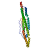 4xxxS S: Starting model for refinement |
|---|---|
| Similar structure data |
-Links
 PDBj
PDBj
- Assembly
| Deposited unit | 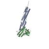
| ||||||||
|---|---|---|---|---|---|---|---|---|---|
| 1 |
| ||||||||
| Unit cell |
|
-Components
| #1: Protein | Mass: 32711.186 Da / Num. of mol.: 1 Source method: isolated from a genetically manipulated source Source: (gene. exp.) Mycobacterium tuberculosis (bacteria) / Strain: ATCC 25618 / H37Rv / Gene: espB, mtb48, Rv3881c, MTV027.16c / Production host: Escherichia coli (E. coli) / References: UniProt: P9WJD9 |
|---|---|
| #2: Protein | Mass: 26447.820 Da / Num. of mol.: 1 Source method: isolated from a genetically manipulated source Source: (gene. exp.) Mycobacterium tuberculosis (bacteria) / Strain: ATCC 25618 / H37Rv / Gene: espK, Rv3879c / Production host: Escherichia coli (E. coli) / References: UniProt: P9WJC1 |
| #3: Water | ChemComp-HOH / Water Mass: 18.015 Da / Num. of mol.: 38 / Source method: isolated from a natural source / Formula: H2O Mass: 18.015 Da / Num. of mol.: 38 / Source method: isolated from a natural source / Formula: H2O |
-Experimental details
-Experiment
| Experiment | Method: X-RAY DIFFRACTION / Number of used crystals: 1 |
|---|
- Sample preparation
| Crystal | Density Matthews: 5.06 Å3/Da / Density % sol: 75.5 % |
|---|---|
| Crystal grow | Temperature: 277 K / Method: vapor diffusion, hanging drop / pH: 7 Details: 0.2-1.2 M sodium malonate 0.1 M HEPES (pH 7.8) 8-14% PEG3350 |
-Data collection
| Diffraction | Mean temperature: 100 K / Serial crystal experiment: N |
|---|---|
| Diffraction source | Source: SYNCHROTRON / Site: ESRF / Beamline: ID30B / Wavelength: 0.976 Å |
| Detector | Type: DECTRIS PILATUS3 6M / Detector: PIXEL / Date: Feb 12, 2021 |
| Radiation | Monochromator: Si / Protocol: SINGLE WAVELENGTH / Monochromatic (M) / Laue (L): M / Scattering type: x-ray |
| Radiation wavelength | Wavelength: 0.976 Å / Relative weight: 1 |
| Reflection | Resolution: 2.29→85.82 Å / Num. obs: 28129 / % possible obs: 53.2 % / Redundancy: 6.2 % / Biso Wilson estimate: 63.86 Å2 / Rpim(I) all: 0.034 / Net I/σ(I): 11.7 |
| Reflection shell | Resolution: 2.3→2.6 Å / Mean I/σ(I) obs: 1.9 / Num. unique obs: 1406 / Rpim(I) all: 0.38 |
- Processing
| Software |
| |||||||||||||||||||||||||||||||||||||||||||||||||||||||||||||||||||||||||||||||||||||||||||||||||||||||||||||||||||||||||||||||||||||||||||||||||||
|---|---|---|---|---|---|---|---|---|---|---|---|---|---|---|---|---|---|---|---|---|---|---|---|---|---|---|---|---|---|---|---|---|---|---|---|---|---|---|---|---|---|---|---|---|---|---|---|---|---|---|---|---|---|---|---|---|---|---|---|---|---|---|---|---|---|---|---|---|---|---|---|---|---|---|---|---|---|---|---|---|---|---|---|---|---|---|---|---|---|---|---|---|---|---|---|---|---|---|---|---|---|---|---|---|---|---|---|---|---|---|---|---|---|---|---|---|---|---|---|---|---|---|---|---|---|---|---|---|---|---|---|---|---|---|---|---|---|---|---|---|---|---|---|---|---|---|---|---|
| Refinement | Method to determine structure: MOLECULAR REPLACEMENT Starting model: 4XXX Resolution: 2.293→85.816 Å / Cor.coef. Fo:Fc: 0.933 / Cor.coef. Fo:Fc free: 0.91 / WRfactor Rfree: 0.28 / WRfactor Rwork: 0.247 / Average fsc free: 0.8555 / Average fsc work: 0.8707 / Cross valid method: THROUGHOUT / ESU R Free: 0.308 Details: Hydrogens have been used if present in the input file
| |||||||||||||||||||||||||||||||||||||||||||||||||||||||||||||||||||||||||||||||||||||||||||||||||||||||||||||||||||||||||||||||||||||||||||||||||||
| Solvent computation | Ion probe radii: 0.8 Å / Shrinkage radii: 0.8 Å / VDW probe radii: 1.2 Å / Solvent model: MASK BULK SOLVENT | |||||||||||||||||||||||||||||||||||||||||||||||||||||||||||||||||||||||||||||||||||||||||||||||||||||||||||||||||||||||||||||||||||||||||||||||||||
| Displacement parameters | Biso mean: 84.479 Å2
| |||||||||||||||||||||||||||||||||||||||||||||||||||||||||||||||||||||||||||||||||||||||||||||||||||||||||||||||||||||||||||||||||||||||||||||||||||
| Refinement step | Cycle: LAST / Resolution: 2.293→85.816 Å
| |||||||||||||||||||||||||||||||||||||||||||||||||||||||||||||||||||||||||||||||||||||||||||||||||||||||||||||||||||||||||||||||||||||||||||||||||||
| Refine LS restraints |
| |||||||||||||||||||||||||||||||||||||||||||||||||||||||||||||||||||||||||||||||||||||||||||||||||||||||||||||||||||||||||||||||||||||||||||||||||||
| LS refinement shell |
|
