 Movie
Movie Controller

[English] 日本語
 Yorodumi
- PDB-8aip: Crystal Structure of Two-domain bacterial laccase from the actino... -
+ Open data

- Basic information
| Entry | Database: PDB / ID: 8aip | ||||||
|---|---|---|---|---|---|---|---|
| Title | Crystal Structure of Two-domain bacterial laccase from the actinobacterium Streptomyces carpinensis VKM Ac-1300 | ||||||
 Components Components | Two-Domain Laccase | ||||||
 Keywords Keywords |  OXIDOREDUCTASE / Two-Domain Laccase / laccase / Streptomyces carpinensis OXIDOREDUCTASE / Two-Domain Laccase / laccase / Streptomyces carpinensis | ||||||
| Function / homology | COPPER (II) ION / OXYGEN MOLECULE Function and homology information Function and homology information | ||||||
| Biological species |  Streptomyces carpinensis (bacteria) Streptomyces carpinensis (bacteria) | ||||||
| Method | X-RAY DIFFRACTION / MOLECULAR REPLACEMENT / Resolution: 2.35 Å | ||||||
 Authors Authors | Gabdulkhakov, A.G. / Tishchenko, T.V. / Trubitsina, L. / Trubitsin, I. / Leontievsky, A. / Lisov, A. | ||||||
| Funding support |  Russian Federation, 1items Russian Federation, 1items
| ||||||
 Citation Citation | Journal: Biochemistry Mosc. / Year: 2023 Title: A Novel Two-Domain Laccase with Middle Redox Potential: Physicochemical and Structural Properties. Authors: Trubitsina, L.I. / Trubitsin, I.V. / Lisov, A.V. / Gabdulkhakov, A.G. / Zavarzina, A.G. / Belova, O.V. / Larionova, A.P. / Tishchenko, S.V. / Leontievsky, A.A. | ||||||
| History |
|
- Structure visualization
| Structure viewer | Molecule: MolmilJmol/JSmol |
|---|
- Downloads & links
-Download
| PDBx/mmCIF format | 8aip.cif.gz | 144.2 KB | Display | PDBx/mmCIF format |
|---|---|---|---|---|
| PDB format | pdb8aip.ent.gz | 92.9 KB | Display | PDB format |
| PDBx/mmJSON format | 8aip.json.gz | Tree view | PDBx/mmJSON format | |
| Others |  Other downloads Other downloads |
-Validation report
| Arichive directory | https://data.pdbj.org/pub/pdb/validation_reports/ai/8aipftp://data.pdbj.org/pub/pdb/validation_reports/ai/8aip | HTTPS FTP |
|---|
-Related structure data
| Related structure data | 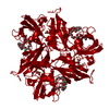 4gybS S: Starting model for refinement |
|---|---|
| Similar structure data |
-Links
 PDBj
PDBj


- Assembly
| Deposited unit | 
| ||||||||||||
|---|---|---|---|---|---|---|---|---|---|---|---|---|---|
| 1 |
| ||||||||||||
| Unit cell |
| ||||||||||||
| Components on special symmetry positions |
|
-Components
| #1: Protein | Mass: 32226.031 Da / Num. of mol.: 1 Source method: isolated from a genetically manipulated source Details: VKM Ac-1300 / Source: (gene. exp.) Streptomyces carpinensis (bacteria) / Production host: Escherichia coli (E. coli) | ||||||
|---|---|---|---|---|---|---|---|
| #2: Chemical | Copper  Mass: 63.546 Da / Num. of mol.: 3 / Source method: obtained synthetically / Formula: Cu / Feature type: SUBJECT OF INVESTIGATION Mass: 63.546 Da / Num. of mol.: 3 / Source method: obtained synthetically / Formula: Cu / Feature type: SUBJECT OF INVESTIGATION#3: Chemical | ChemComp-OXY / | Oxygen  Mass: 31.999 Da / Num. of mol.: 1 / Source method: obtained synthetically / Formula: O2 / Feature type: SUBJECT OF INVESTIGATION Mass: 31.999 Da / Num. of mol.: 1 / Source method: obtained synthetically / Formula: O2 / Feature type: SUBJECT OF INVESTIGATION#4: Water | ChemComp-HOH / | Water Mass: 18.015 Da / Num. of mol.: 83 / Source method: isolated from a natural source / Formula: H2O Mass: 18.015 Da / Num. of mol.: 83 / Source method: isolated from a natural source / Formula: H2OHas ligand of interest | Y | |
-Experimental details
-Experiment
| Experiment | Method: X-RAY DIFFRACTION / Number of used crystals: 1 |
|---|
- Sample preparation
| Crystal | Density Matthews: 2.43 Å3/Da / Density % sol: 49.4 % |
|---|---|
| Crystal grow | Temperature: 297 K / Method: vapor diffusion, hanging drop Details: 20%v/v PEG Smear Broad, 0.1 M Sodium citratate, pH 5.6, 0.15 M Mg acetate (condition #33 of BCS-1 from Molecular Dimensions, UK) |
-Data collection
| Diffraction | Mean temperature: 110 K / Serial crystal experiment: N |
|---|---|
| Diffraction source | Source: ROTATING ANODE / Type: BRUKER X8 PROTEUM / Wavelength: 1.54178 Å |
| Detector | Type: Bruker Platinum 135 / Detector: CCD / Date: Jan 30, 2020 |
| Radiation | Protocol: SINGLE WAVELENGTH / Monochromatic (M) / Laue (L): M / Scattering type: x-ray |
| Radiation wavelength | Wavelength: 1.54178 Å / Relative weight: 1 |
| Reflection | Resolution: 2.3→33 Å / Num. obs: 13826 / % possible obs: 97 % / Redundancy: 4.29 % / Biso Wilson estimate: 30.39 Å2 / CC1/2: 0.99 / Rmerge(I) obs: 0.075 / Net I/σ(I): 6.8 |
| Reflection shell | Resolution: 2.3→2.38 Å / Rmerge(I) obs: 0.355 / Mean I/σ(I) obs: 1.5 / Num. unique obs: 1147 / CC1/2: 0.8 / % possible all: 82.3 |
- Processing
| Software |
| ||||||||||||||||||||||||||||||||||||||||||
|---|---|---|---|---|---|---|---|---|---|---|---|---|---|---|---|---|---|---|---|---|---|---|---|---|---|---|---|---|---|---|---|---|---|---|---|---|---|---|---|---|---|---|---|
| Refinement | Method to determine structure: MOLECULAR REPLACEMENT Starting model: 4GYB Resolution: 2.35→32.65 Å / SU ML: 0.3312 / Cross valid method: FREE R-VALUE / σ(F): 1.33 / Phase error: 33.6689 Stereochemistry target values: GeoStd + Monomer Library + CDL v1.2
| ||||||||||||||||||||||||||||||||||||||||||
| Solvent computation | Shrinkage radii: 0.9 Å / VDW probe radii: 1.11 Å / Solvent model: FLAT BULK SOLVENT MODEL | ||||||||||||||||||||||||||||||||||||||||||
| Displacement parameters | Biso mean: 30.3 Å2 | ||||||||||||||||||||||||||||||||||||||||||
| Refinement step | Cycle: LAST / Resolution: 2.35→32.65 Å
| ||||||||||||||||||||||||||||||||||||||||||
| Refine LS restraints |
| ||||||||||||||||||||||||||||||||||||||||||
| LS refinement shell |
| ||||||||||||||||||||||||||||||||||||||||||
| Refinement TLS params. | Method: refined / Origin x: 38.8202093805 Å / Origin y: -15.2701824309 Å / Origin z: 20.310484471 Å
| ||||||||||||||||||||||||||||||||||||||||||
| Refinement TLS group | Selection details: all |
