 Movie
Movie Controller


+ Open data

- Basic information
| Entry | Database: PDB / ID: 8agr | |||||||||
|---|---|---|---|---|---|---|---|---|---|---|
| Title | Crystal structure of DltE from L. plantarum, apo form | |||||||||
 Components Components | Beta-lactamase family protein | |||||||||
 Keywords Keywords |  CELL CYCLE / carboxyesterase / D-alanylation / lipoteichoic acids CELL CYCLE / carboxyesterase / D-alanylation / lipoteichoic acids | |||||||||
| Function / homology | serine-type D-Ala-D-Ala carboxypeptidase / serine-type D-Ala-D-Ala carboxypeptidase activity / Beta-lactamase-related / Beta-lactamase / Beta-lactamase/transpeptidase-like / Serine-type D-Ala-D-Ala carboxypeptidase Function and homology information Function and homology information | |||||||||
| Biological species |  Lactiplantibacillus plantarum (bacteria) Lactiplantibacillus plantarum (bacteria) | |||||||||
| Method | X-RAY DIFFRACTION / SYNCHROTRON / MOLECULAR REPLACEMENT / Resolution: 1.86 Å | |||||||||
 Authors Authors | Ravaud, S. / Nikolopoulos, N. / Grangeasse, C. | |||||||||
| Funding support |  France, 2items France, 2items
| |||||||||
 Citation Citation | Journal: Elife / Year: 2023 Title: Structure-function analysis of Lactiplantibacillus plantarum DltE& reveals D-alanylated lipoteichoic acids as direct cues supporting Drosophila juvenile growth. Authors: Nikolopoulos, N. / Matos, R. / Ravaud, S. / Courtin, P. / Akherraz, H. / Palussiere, S. / Gueguen-Chaignon, V. / Salomon-Mallet, M. / Guillot, A. / Guerardel, Y. / Chapot-Chartier, M.P. / ...Authors: Nikolopoulos, N. / Matos, R. / Ravaud, S. / Courtin, P. / Akherraz, H. / Palussiere, S. / Gueguen-Chaignon, V. / Salomon-Mallet, M. / Guillot, A. / Guerardel, Y. / Chapot-Chartier, M.P. / Grangeasse, C. / Leulier, F. | |||||||||
| History |
|
- Structure visualization
| Structure viewer | Molecule: MolmilJmol/JSmol |
|---|
- Downloads & links
-Download
| PDBx/mmCIF format | 8agr.cif.gz | 165.7 KB | Display | PDBx/mmCIF format |
|---|---|---|---|---|
| PDB format | pdb8agr.ent.gz | 124.2 KB | Display | PDB format |
| PDBx/mmJSON format | 8agr.json.gz | Tree view | PDBx/mmJSON format | |
| Others |  Other downloads Other downloads |
-Validation report
| Arichive directory | https://data.pdbj.org/pub/pdb/validation_reports/ag/8agrftp://data.pdbj.org/pub/pdb/validation_reports/ag/8agr | HTTPS FTP |
|---|
-Related structure data
| Related structure data | 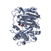 8aikC 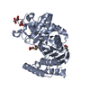 8ajiC 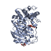 8akhC  3wwxS S: Starting model for refinement C: citing same article ( |
|---|---|
| Similar structure data |
-Links
 PDBj
PDBj
- Assembly
| Deposited unit | 
| ||||||||
|---|---|---|---|---|---|---|---|---|---|
| 1 | 
| ||||||||
| 2 | 
| ||||||||
| Unit cell |
|
-Components
| #1: Protein | Mass: 41822.508 Da / Num. of mol.: 2 Source method: isolated from a genetically manipulated source Source: (gene. exp.) Lactiplantibacillus plantarum (bacteria)Gene: pbpX2, pbpX_4, C7M36_02596, E3O64_07015, E3U93_06380, FEE41_04205, IV39_GL001648, LPJSA22_01868, SN35N_2987 Production host: Escherichia coli (E. coli)References: UniProt: A0A0P7JVD2, serine-type D-Ala-D-Ala carboxypeptidase#2: Chemical | Sulfate  Mass: 96.063 Da / Num. of mol.: 3 / Source method: obtained synthetically / Formula: SO4 Mass: 96.063 Da / Num. of mol.: 3 / Source method: obtained synthetically / Formula: SO4#3: Chemical | ChemComp-EDO / Ethylene glycol  Mass: 62.068 Da / Num. of mol.: 4 / Source method: obtained synthetically / Formula: C2H6O2 Mass: 62.068 Da / Num. of mol.: 4 / Source method: obtained synthetically / Formula: C2H6O2#4: Water | ChemComp-HOH / | Water Mass: 18.015 Da / Num. of mol.: 845 / Source method: isolated from a natural source / Formula: H2O Mass: 18.015 Da / Num. of mol.: 845 / Source method: isolated from a natural source / Formula: H2OHas ligand of interest | N | |
|---|
-Experimental details
-Experiment
| Experiment | Method: X-RAY DIFFRACTION / Number of used crystals: 1 |
|---|
- Sample preparation
| Crystal | Density Matthews: 2.35 Å3/Da / Density % sol: 47.77 % |
|---|---|
| Crystal grow | Temperature: 293 K / Method: vapor diffusion, sitting drop / Details: PEG MME 5K, Tris pH8.5, LiSO4, ethylene glycol |
-Data collection
| Diffraction | Mean temperature: 100 K / Serial crystal experiment: N |
|---|---|
| Diffraction source | Source: SYNCHROTRON / Site: ESRF / Beamline: ID23-2 / Wavelength: 0.8731 Å |
| Detector | Type: DECTRIS PILATUS3 2M / Detector: PIXEL / Date: Apr 18, 2018 |
| Radiation | Protocol: SINGLE WAVELENGTH / Monochromatic (M) / Laue (L): M / Scattering type: x-ray |
| Radiation wavelength | Wavelength: 0.8731 Å / Relative weight: 1 |
| Reflection | Resolution: 1.86→47.86 Å / Num. obs: 64951 / % possible obs: 99.9 % / Redundancy: 4.5 % / Rpim(I) all: 0.12 / Net I/σ(I): 6.4 |
| Reflection shell | Resolution: 1.86→1.9 Å / Mean I/σ(I) obs: 2 / Num. unique obs: 39993 / Rpim(I) all: 0.441 |
- Processing
| Software |
| |||||||||||||||||||||||||||||||||||||||||||||||||||||||||||||||||||||||||||||||||||||||||||||||||||||||||||||||||||||||||||||||||||||||||||||||||||||||||||||||||||||||||||||||||||||||||||||||||||||||||||||||||||||||||||||||||||||||
|---|---|---|---|---|---|---|---|---|---|---|---|---|---|---|---|---|---|---|---|---|---|---|---|---|---|---|---|---|---|---|---|---|---|---|---|---|---|---|---|---|---|---|---|---|---|---|---|---|---|---|---|---|---|---|---|---|---|---|---|---|---|---|---|---|---|---|---|---|---|---|---|---|---|---|---|---|---|---|---|---|---|---|---|---|---|---|---|---|---|---|---|---|---|---|---|---|---|---|---|---|---|---|---|---|---|---|---|---|---|---|---|---|---|---|---|---|---|---|---|---|---|---|---|---|---|---|---|---|---|---|---|---|---|---|---|---|---|---|---|---|---|---|---|---|---|---|---|---|---|---|---|---|---|---|---|---|---|---|---|---|---|---|---|---|---|---|---|---|---|---|---|---|---|---|---|---|---|---|---|---|---|---|---|---|---|---|---|---|---|---|---|---|---|---|---|---|---|---|---|---|---|---|---|---|---|---|---|---|---|---|---|---|---|---|---|---|---|---|---|---|---|---|---|---|---|---|---|---|---|---|---|---|
| Refinement | Method to determine structure: MOLECULAR REPLACEMENT Starting model: 3WWX Resolution: 1.86→47.796 Å / Cor.coef. Fo:Fc: 0.955 / Cor.coef. Fo:Fc free: 0.941 / WRfactor Rfree: 0.202 / WRfactor Rwork: 0.165 / SU B: 2.901 / SU ML: 0.086 / Average fsc free: 0.9732 / Average fsc work: 0.9822 / Cross valid method: FREE R-VALUE / ESU R: 0.126 / ESU R Free: 0.12 Details: Hydrogens have been added in their riding positions
| |||||||||||||||||||||||||||||||||||||||||||||||||||||||||||||||||||||||||||||||||||||||||||||||||||||||||||||||||||||||||||||||||||||||||||||||||||||||||||||||||||||||||||||||||||||||||||||||||||||||||||||||||||||||||||||||||||||||
| Solvent computation | Ion probe radii: 0.8 Å / Shrinkage radii: 0.8 Å / VDW probe radii: 1.2 Å / Solvent model: MASK BULK SOLVENT | |||||||||||||||||||||||||||||||||||||||||||||||||||||||||||||||||||||||||||||||||||||||||||||||||||||||||||||||||||||||||||||||||||||||||||||||||||||||||||||||||||||||||||||||||||||||||||||||||||||||||||||||||||||||||||||||||||||||
| Displacement parameters | Biso mean: 14.051 Å2
| |||||||||||||||||||||||||||||||||||||||||||||||||||||||||||||||||||||||||||||||||||||||||||||||||||||||||||||||||||||||||||||||||||||||||||||||||||||||||||||||||||||||||||||||||||||||||||||||||||||||||||||||||||||||||||||||||||||||
| Refinement step | Cycle: LAST / Resolution: 1.86→47.796 Å
| |||||||||||||||||||||||||||||||||||||||||||||||||||||||||||||||||||||||||||||||||||||||||||||||||||||||||||||||||||||||||||||||||||||||||||||||||||||||||||||||||||||||||||||||||||||||||||||||||||||||||||||||||||||||||||||||||||||||
| Refine LS restraints |
| |||||||||||||||||||||||||||||||||||||||||||||||||||||||||||||||||||||||||||||||||||||||||||||||||||||||||||||||||||||||||||||||||||||||||||||||||||||||||||||||||||||||||||||||||||||||||||||||||||||||||||||||||||||||||||||||||||||||
| LS refinement shell | Refine-ID: X-RAY DIFFRACTION / Total num. of bins used: 20
|
