 Movie
Movie Controller

[English] 日本語
 Yorodumi
- PDB-8af4: Room temperature SSX structure of GH11 xylanase from Nectria haem... -
+ Open data

- Basic information
| Entry | Database: PDB / ID: 8af4 | ||||||
|---|---|---|---|---|---|---|---|
| Title | Room temperature SSX structure of GH11 xylanase from Nectria haematococca (40000 frames) | ||||||
 Components Components | Endo-1,4-beta-xylanase Xylanase Xylanase | ||||||
 Keywords Keywords | HYDROLASE / Xylanase / SSX / TapeDrive / room temperature | ||||||
| Function / homology |  Function and homology informationendo-1,4-beta-xylanase activity / endo-1,4-beta-xylanase / xylan catabolic process Function and homology informationendo-1,4-beta-xylanase activity / endo-1,4-beta-xylanase / xylan catabolic processSimilarity search - Function | ||||||
| Biological species |  Fusarium haematococcum (fungus) Fusarium haematococcum (fungus) | ||||||
| Method | X-RAY DIFFRACTION / SYNCHROTRON / MOLECULAR REPLACEMENT / Resolution: 1.51 Å | ||||||
 Authors Authors | Oberthuer, D. / Andaleeb, H. / Betzel, C. / Perbandt, M. / Yefanov, O. / Zielinski, K. | ||||||
| Funding support | 1items
| ||||||
 Citation Citation | Journal: Iucrj / Year: 2022 Title: Rapid and efficient room-temperature serial synchrotron crystallography using the CFEL TapeDrive. Authors: Zielinski, K.A. / Prester, A. / Andaleeb, H. / Bui, S. / Yefanov, O. / Catapano, L. / Henkel, A. / Wiedorn, M.O. / Lorbeer, O. / Crosas, E. / Meyer, J. / Mariani, V. / Domaracky, M. / White, ...Authors: Zielinski, K.A. / Prester, A. / Andaleeb, H. / Bui, S. / Yefanov, O. / Catapano, L. / Henkel, A. / Wiedorn, M.O. / Lorbeer, O. / Crosas, E. / Meyer, J. / Mariani, V. / Domaracky, M. / White, T.A. / Fleckenstein, H. / Sarrou, I. / Werner, N. / Betzel, C. / Rohde, H. / Aepfelbacher, M. / Chapman, H.N. / Perbandt, M. / Steiner, R.A. / Oberthuer, D. | ||||||
| History |
|
- Structure visualization
| Structure viewer | Molecule: MolmilJmol/JSmol |
|---|
- Downloads & links
-Download
| PDBx/mmCIF format | 8af4.cif.gz | 110 KB | Display | PDBx/mmCIF format |
|---|---|---|---|---|
| PDB format | pdb8af4.ent.gz | 69.1 KB | Display | PDB format |
| PDBx/mmJSON format | 8af4.json.gz | Tree view | PDBx/mmJSON format | |
| Others |  Other downloads Other downloads |
-Validation report
| Arichive directory | https://data.pdbj.org/pub/pdb/validation_reports/af/8af4ftp://data.pdbj.org/pub/pdb/validation_reports/af/8af4 | HTTPS FTP |
|---|
-Related structure data
| Related structure data | 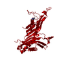 7qarC 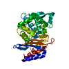 7zpvC 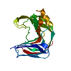 7zq0C 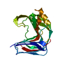 8af5C  8af6C 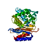 8af7C 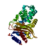 8af8C 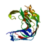 6y0hS S: Starting model for refinement C: citing same article ( |
|---|---|
| Similar structure data |
-Links
 PDBj
PDBj
- Assembly
| Deposited unit | 
| ||||||||||||
|---|---|---|---|---|---|---|---|---|---|---|---|---|---|
| 1 |
| ||||||||||||
| Unit cell |
| ||||||||||||
| Components on special symmetry positions |
|
-Components
| #1: Protein | Xylanase Mass: 20575.037 Da / Num. of mol.: 1 Source method: isolated from a genetically manipulated source Source: (gene. exp.) Fusarium haematococcum (fungus)Strain: ATCC MYA-4622 / CBS 123669 / FGSC 9596 / NRRL 45880 / 77-13-4 Gene: NECHADRAFT_106153 / Production host:  Escherichia coli BL21(DE3) (bacteria) / References: UniProt: C7YSL3, endo-1,4-beta-xylanase Escherichia coli BL21(DE3) (bacteria) / References: UniProt: C7YSL3, endo-1,4-beta-xylanase |
|---|---|
| #2: Water | ChemComp-HOH / Water Mass: 18.015 Da / Num. of mol.: 220 / Source method: isolated from a natural source / Formula: H2O Mass: 18.015 Da / Num. of mol.: 220 / Source method: isolated from a natural source / Formula: H2O |
-Experimental details
-Experiment
| Experiment | Method: X-RAY DIFFRACTION / Number of used crystals: 1 |
|---|
- Sample preparation
| Crystal | Density Matthews: 2.04 Å3/Da / Density % sol: 39.61 % |
|---|---|
| Crystal grow | Temperature: 293 K / Method: batch mode / pH: 5.5 Details: For crystallization, the original conditions were modified to obtain microcrystals. Initial crystals obtained from hanging drops under the precipitant condition: 1 M ammonium sulfate, 100 mM ...Details: For crystallization, the original conditions were modified to obtain microcrystals. Initial crystals obtained from hanging drops under the precipitant condition: 1 M ammonium sulfate, 100 mM sodium citrate pH 5.5 were crushed under a stereomicroscope, using a crystal crusher tool (Hampton research). The reservoir solution was pipetted to the drop and the seed stock was collected by washing the drop with reservoir solution. The seed stock was transferred to a seed bead tube (Molecular Dimensions Ltd., UK), vortexed three times for 30 s each, with an interval of 30 s between each vortex, to get the final seed stock. Protein solution 15 mg/mL, precipitant solution, and seed stock were mixed with a ratio of 1:1:0.5. The mixture was vortexed for 30 s in ten-minute intervals. After 30 min, the microcrystals were centrifuged at 200 rpm and the supernatant was replaced with a precipitant solution. Applying the same protocol, microcrystals were obtained under two precipitant conditions i.e. Precipitant 1: 1M (NH4)2SO4, 100 mM sodium citrate pH 5.5, and Precipitant 2: 200 mM (NH4)2SO4, 100 mM sodium citrate pH 5.5 and 20% PEG 6000. Microcrystals obtained under both precipitant conditions were tested for diffraction data collection |
-Data collection
| Diffraction | Mean temperature: 295 K / Serial crystal experiment: Y |
|---|---|
| Diffraction source | Source: SYNCHROTRON / Site: PETRA III, DESY  / Beamline: P11 / Wavelength: 1.0332 Å / Beamline: P11 / Wavelength: 1.0332 Å |
| Detector | Type: DECTRIS PILATUS 6M / Detector: PIXEL / Date: Dec 10, 2019 |
| Radiation | Protocol: SINGLE WAVELENGTH / Monochromatic (M) / Laue (L): M / Scattering type: x-ray |
| Radiation wavelength | Wavelength: 1.0332 Å / Relative weight: 1 |
| Reflection | Resolution: 1.51→17.16 Å / Num. obs: 25916 / % possible obs: 98.75 % / Redundancy: 432 % / Biso Wilson estimate: 19.51 Å2 / CC1/2: 0.987 / CC star: 0.997 / R split: 0.1342 / Net I/σ(I): 5.83 |
| Reflection shell | Resolution: 1.51→1.54 Å / Mean I/σ(I) obs: 0.55 / Num. unique obs: 2291 / CC1/2: 0.152 / CC star: 0.514 / R split: 2.49 / % possible all: 87.48 |
| Serial crystallography sample delivery | Description: TapeDrive / Method: injection |
| Serial crystallography sample delivery injection | Description: TapeDrive / Flow rate: 1 µL/min / Injector diameter: 180 µm / Injector temperature: 295 K |
| Serial crystallography data reduction | Frames total: 40000 / Lattices indexed: 41508 |
- Processing
| Software |
| |||||||||||||||||||||||||||||||||||||||||||||||||||||||||||||||||||||||||||||||||||||||||||||||||||||||||
|---|---|---|---|---|---|---|---|---|---|---|---|---|---|---|---|---|---|---|---|---|---|---|---|---|---|---|---|---|---|---|---|---|---|---|---|---|---|---|---|---|---|---|---|---|---|---|---|---|---|---|---|---|---|---|---|---|---|---|---|---|---|---|---|---|---|---|---|---|---|---|---|---|---|---|---|---|---|---|---|---|---|---|---|---|---|---|---|---|---|---|---|---|---|---|---|---|---|---|---|---|---|---|---|---|---|---|
| Refinement | Method to determine structure: MOLECULAR REPLACEMENT Starting model: 6Y0H Resolution: 1.51→17.16 Å / SU ML: 0.1996 / Cross valid method: FREE R-VALUE / σ(F): 1.33 / Phase error: 21.4836 Stereochemistry target values: GeoStd + Monomer Library + CDL v1.2
| |||||||||||||||||||||||||||||||||||||||||||||||||||||||||||||||||||||||||||||||||||||||||||||||||||||||||
| Solvent computation | Shrinkage radii: 0.9 Å / VDW probe radii: 1.11 Å / Solvent model: FLAT BULK SOLVENT MODEL | |||||||||||||||||||||||||||||||||||||||||||||||||||||||||||||||||||||||||||||||||||||||||||||||||||||||||
| Displacement parameters | Biso mean: 23.58 Å2 | |||||||||||||||||||||||||||||||||||||||||||||||||||||||||||||||||||||||||||||||||||||||||||||||||||||||||
| Refinement step | Cycle: LAST / Resolution: 1.51→17.16 Å
| |||||||||||||||||||||||||||||||||||||||||||||||||||||||||||||||||||||||||||||||||||||||||||||||||||||||||
| Refine LS restraints |
| |||||||||||||||||||||||||||||||||||||||||||||||||||||||||||||||||||||||||||||||||||||||||||||||||||||||||
| LS refinement shell |
|
