 Movie
Movie Controller

[English] 日本語
 Yorodumi
- PDB-8a8c: T5 phage receptor-binding protein pb5 bound to ferrichrome transp... -
+ Open data

- Basic information
| Entry | Database: PDB / ID: 8a8c | ||||||
|---|---|---|---|---|---|---|---|
| Title | T5 phage receptor-binding protein pb5 bound to ferrichrome transporter FhuA | ||||||
 Components Components |
| ||||||
 Keywords Keywords |  MEMBRANE PROTEIN / Bacteriophage / receptor / TonB-dependent transporter / outer membrane MEMBRANE PROTEIN / Bacteriophage / receptor / TonB-dependent transporter / outer membrane | ||||||
| Function / homology |  Function and homology information Function and homology informationsiderophore transmembrane transport / siderophore uptake transmembrane transporter activity / virus tail / transmembrane transporter complex / virion binding / toxic substance binding / cell outer membrane / signaling receptor activity / intracellular iron ion homeostasis / entry receptor-mediated virion attachment to host cell ...siderophore transmembrane transport / siderophore uptake transmembrane transporter activity / virus tail / transmembrane transporter complex / virion binding / toxic substance binding / cell outer membrane / signaling receptor activity / intracellular iron ion homeostasis / entry receptor-mediated virion attachment to host cell / receptor-mediated virion attachment to host cell / symbiont entry into host cell / iron ion binding / protein domain specific binding / membraneSimilarity search - Function | ||||||
| Biological species |  Escherichia coli K-12 (bacteria) Escherichia coli K-12 (bacteria) Escherichia phage T5 (virus) Escherichia phage T5 (virus) | ||||||
| Method | ELECTRON MICROSCOPY / single particle reconstruction / cryo EM / Resolution: 3.1 Å | ||||||
 Authors Authors | Silale, A. / van den Berg, B. | ||||||
| Funding support |  United Kingdom, 1items United Kingdom, 1items
| ||||||
 Citation Citation | Journal: Proc Natl Acad Sci U S A / Year: 2022 Title: Structural basis for host recognition and superinfection exclusion by bacteriophage T5. Authors: Bert van den Berg / Augustinas Silale / Arnaud Baslé / Astrid F Brandner / Sophie L Mader / Syma Khalid / Abstract: A key but poorly understood stage of the bacteriophage life cycle is the binding of phage receptor-binding proteins (RBPs) to receptors on the host cell surface, leading to injection of the phage ...A key but poorly understood stage of the bacteriophage life cycle is the binding of phage receptor-binding proteins (RBPs) to receptors on the host cell surface, leading to injection of the phage genome and, for lytic phages, host cell lysis. To prevent secondary infection by the same or a closely related phage and nonproductive phage adsorption to lysed cell fragments, superinfection exclusion (SE) proteins can prevent the binding of RBPs via modulation of the host receptor structure in ways that are also unclear. Here, we present the cryogenic electron microscopy (cryo-EM) structure of the phage T5 outer membrane (OM) receptor FhuA in complex with the T5 RBP pb5, and the crystal structure of FhuA complexed to the OM SE lipoprotein Llp. Pb5 inserts four loops deeply into the extracellular lumen of FhuA and contacts the plug but does not cause any conformational changes in the receptor, supporting the view that DNA translocation does not occur through the lumen of OM channels. The FhuA-Llp structure reveals that Llp is periplasmic and binds to a nonnative conformation of the plug of FhuA, causing the inward folding of two extracellular loops via "reverse" allostery. The inward-folded loops of FhuA overlap with the pb5 binding site, explaining how Llp binding to FhuA abolishes further infection of by phage T5 and suggesting a mechanism for SE via the jamming of TonB-dependent transporters by small phage lipoproteins. | ||||||
| History |
|
- Structure visualization
| Structure viewer | Molecule: MolmilJmol/JSmol |
|---|
- Downloads & links
-Download
| PDBx/mmCIF format | 8a8c.cif.gz | 242.9 KB | Display | PDBx/mmCIF format |
|---|---|---|---|---|
| PDB format | pdb8a8c.ent.gz | 195.5 KB | Display | PDB format |
| PDBx/mmJSON format | 8a8c.json.gz | Tree view | PDBx/mmJSON format | |
| Others |  Other downloads Other downloads |
-Validation report
| Arichive directory | https://data.pdbj.org/pub/pdb/validation_reports/a8/8a8cftp://data.pdbj.org/pub/pdb/validation_reports/a8/8a8c | HTTPS FTP |
|---|
-Related structure data
| Related structure data |  15229MC  8a60C M: map data used to model this data C: citing same article ( |
|---|---|
| Similar structure data |
-Links
 PDBj
PDBj- Assembly
| Deposited unit | 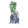
|
|---|---|
| 1 |
|
-Components
| #1: Protein | Mass: 78816.859 Da / Num. of mol.: 1 Source method: isolated from a genetically manipulated source Source: (gene. exp.) Escherichia coli K-12 (bacteria) / Strain: K12 / Gene: fhuA, tonA, b0150, JW0146 / Production host: Escherichia coli (E. coli) / References: UniProt: P06971 |
|---|---|
| #2: Protein | Mass: 69611.453 Da / Num. of mol.: 1 Source method: isolated from a genetically manipulated source Source: (gene. exp.) Escherichia phage T5 (virus) / Gene: oad, T5.157, T5p153 / Production host: Escherichia coli (E. coli) / References: UniProt: P23207 |
| #3: Chemical | ChemComp-LU9 / [(Lipopolysaccharide  Mass: 1996.235 Da / Num. of mol.: 1 / Source method: obtained synthetically / Formula: C93H164N2O39P2 / Comment: toxin*YM Mass: 1996.235 Da / Num. of mol.: 1 / Source method: obtained synthetically / Formula: C93H164N2O39P2 / Comment: toxin*YM |
| Has ligand of interest | N |
-Experimental details
-Experiment
| Experiment | Method: ELECTRON MICROSCOPY |
|---|---|
| EM experiment | Aggregation state: PARTICLE / 3D reconstruction method: single particle reconstruction |
- Sample preparation
| Component | Name: T5 phage receptor-binding protein pb5 bound to ferrichrome transporter FhuA Type: COMPLEX / Entity ID: #1-#2 / Source: RECOMBINANT | ||||||||||||||||||||
|---|---|---|---|---|---|---|---|---|---|---|---|---|---|---|---|---|---|---|---|---|---|
| Molecular weight | Value: 0.148 MDa / Experimental value: NO | ||||||||||||||||||||
| Source (natural) | Organism: Escherichia phage T5 (virus) | ||||||||||||||||||||
| Source (recombinant) | Organism: Escherichia coli (E. coli) | ||||||||||||||||||||
| Buffer solution | pH: 7.5 | ||||||||||||||||||||
| Buffer component |
| ||||||||||||||||||||
| Specimen | Conc.: 8 mg/ml / Embedding applied: NO / Shadowing applied: NO / Staining applied: NO / Vitrification applied: YES | ||||||||||||||||||||
| Specimen support | Grid material: COPPER / Grid mesh size: 300 divisions/in. / Grid type: Quantifoil R1.2/1.3 | ||||||||||||||||||||
| Vitrification | Instrument: FEI VITROBOT MARK IV / Cryogen name: ETHANE / Humidity: 100 % / Chamber temperature: 277 K |
- Electron microscopy imaging
| Experimental equipment |  Model: Titan Krios / Image courtesy: FEI Company |
|---|---|
| Microscopy | Model: FEI TITAN KRIOS |
| Electron gun | Electron source: FIELD EMISSION GUN / Accelerating voltage: 300 kV / Illumination mode: FLOOD BEAM |
| Electron lens | Mode: BRIGHT FIELDBright-field microscopy / Nominal magnification: 165000 X / Nominal defocus max: 2000 nm / Nominal defocus min: 1000 nm / Cs: 2.7 mm / C2 aperture diameter: 50 µm / Alignment procedure: COMA FREE |
| Specimen holder | Cryogen: NITROGEN / Specimen holder model: FEI TITAN KRIOS AUTOGRID HOLDER |
| Image recording | Average exposure time: 2.74 sec. / Electron dose: 40 e/Å2 / Film or detector model: FEI FALCON IV (4k x 4k) / Num. of grids imaged: 1 / Num. of real images: 8387 |
| EM imaging optics | Energyfilter name: TFS Selectris / Energyfilter slit width: 10 eV |
- Processing
| EM software |
| ||||||||||||||||||||||||
|---|---|---|---|---|---|---|---|---|---|---|---|---|---|---|---|---|---|---|---|---|---|---|---|---|---|
| CTF correction | Type: PHASE FLIPPING AND AMPLITUDE CORRECTION | ||||||||||||||||||||||||
| Symmetry | Point symmetry: C1 (asymmetric) | ||||||||||||||||||||||||
| 3D reconstruction | Resolution: 3.1 Å / Resolution method: FSC 0.143 CUT-OFF / Num. of particles: 71476 / Symmetry type: POINT |
