Entry Database : PDB / ID : 7ztlTitle Crystal structure of a covalently linked Aurora-A N-Myc complex Aurora kinase A N-myc proto-oncogene protein Keywords / / Function / homology Function Domain/homology Component
/ / / / / / / / / / / / / / / / / / / / / / / / / / / / / / / / / / / / / / / / / / / / / / / / / / / / / / / / / / / / / / / / / / / / / / / / / / / / / / / / / / / / / / / / / / / / / / / / / / / / / / / / / / / / / / / / / / / / / / / / / / / / / / / / / / / / / / / Biological species Homo sapiens (human)Method / / / Resolution : 1.9 Å Authors Diebold, M. / Schindelin, H. Funding support Organization Grant number Country German Research Foundation (DFG) GRK2243
Journal : Acta Crystallogr D Struct Biol / Year : 2023Title : Crystal structure of a covalently linked Aurora-A-MYCN complex.Authors : Diebold, M. / Schonemann, L. / Eilers, M. / Sotriffer, C. / Schindelin, H. History Deposition May 11, 2022 Deposition site / Processing site Revision 1.0 Jan 18, 2023 Provider / Type Revision 1.1 Jan 31, 2024 Group / Refinement descriptionCategory / chem_comp_bond / pdbx_initial_refinement_modelRevision 1.2 Apr 3, 2024 Group / Category / citation_authorItem _citation.country / _citation.journal_abbrev ... _citation.country / _citation.journal_abbrev / _citation.journal_id_ASTM / _citation.journal_id_ISSN / _citation.pdbx_database_id_DOI / _citation.pdbx_database_id_PubMed / _citation.title / _citation_author.identifier_ORCID
Show all Show less
 Movie
Movie Controller


 Open data
 Basic information
 Components
 Keywords
 TRANSFERASE / Crosslink / protein-protein-complex
 Function and homology information
 Authors
 Germany, 1items
  Citation
 Structure visualization
 Downloads & links
 Other downloads
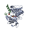
 PDBj
PDBj


 Assembly


 Mass: 427.201 Da / Num. of mol.: 1 / Source method: obtained synthetically / Formula: C10H15N5O10P2 / Comment: ADP, energy-carrying molecule*YM
 Mass: 24.305 Da / Num. of mol.: 2 / Source method: obtained synthetically / Formula: Mg
 Mass: 92.094 Da / Num. of mol.: 3 / Source method: obtained synthetically / Formula: C3H8O3
 Mass: 163.172 Da / Num. of mol.: 1 / Source method: obtained synthetically / Formula: C6H13NO4 / Comment: pH buffer*YM
 Mass: 22.990 Da / Num. of mol.: 1 / Source method: obtained synthetically / Formula: Na
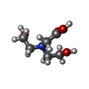
 Mass: 189.166 Da / Num. of mol.: 1 / Source method: obtained synthetically / Formula: C7H11NO5 / Feature type: SUBJECT OF INVESTIGATION
 Sample preparation
 Processing