 Movie
Movie Controller

[English] 日本語
 Yorodumi
- PDB-7zn8: Crystal structure of the light-driven inward proton pump xenorhod... -
+ Open data

- Basic information
| Entry | Database: PDB / ID: 7zn8 | ||||||
|---|---|---|---|---|---|---|---|
| Title | Crystal structure of the light-driven inward proton pump xenorhodopsin BcXeR in the ground state at pH 7.0 in the presence of sodium at 100K | ||||||
 Components Components | xenorhodopsin | ||||||
 Keywords Keywords |  MEMBRANE PROTEIN / retinal / rhodopsin / xenorhodopsin / ion transport / isomerization / photocycle MEMBRANE PROTEIN / retinal / rhodopsin / xenorhodopsin / ion transport / isomerization / photocycle | ||||||
| Function / homology | EICOSANE / OLEIC ACID / (2R)-2,3-dihydroxypropyl (9Z)-octadec-9-enoate / PHOSPHATE ION Function and homology information Function and homology information | ||||||
| Biological species |  | ||||||
| Method | X-RAY DIFFRACTION / SYNCHROTRON / MOLECULAR REPLACEMENT / Resolution: 2.2 Å | ||||||
 Authors Authors | Kovalev, K. / Tsybrov, F. / Alekseev, A. / Bourenkov, G. / Gordeliy, V. | ||||||
| Funding support |  France, 1items France, 1items
| ||||||
 Citation Citation | Journal: Nat.Struct.Mol.Biol. / Year: 2023 Title: Mechanisms of inward transmembrane proton translocation. Authors: Kovalev, K. / Tsybrov, F. / Alekseev, A. / Shevchenko, V. / Soloviov, D. / Siletsky, S. / Bourenkov, G. / Agthe, M. / Nikolova, M. / von Stetten, D. / Astashkin, R. / Bukhdruker, S. / ...Authors: Kovalev, K. / Tsybrov, F. / Alekseev, A. / Shevchenko, V. / Soloviov, D. / Siletsky, S. / Bourenkov, G. / Agthe, M. / Nikolova, M. / von Stetten, D. / Astashkin, R. / Bukhdruker, S. / Chizhov, I. / Royant, A. / Kuzmin, A. / Gushchin, I. / Rosselli, R. / Rodriguez-Valera, F. / Ilyinskiy, N. / Rogachev, A. / Borshchevskiy, V. / Schneider, T.R. / Bamberg, E. / Gordeliy, V. | ||||||
| History |
|
- Structure visualization
| Structure viewer | Molecule: MolmilJmol/JSmol |
|---|
- Downloads & links
-Download
| PDBx/mmCIF format | 7zn8.cif.gz | 175.4 KB | Display | PDBx/mmCIF format |
|---|---|---|---|---|
| PDB format | pdb7zn8.ent.gz | 137.2 KB | Display | PDB format |
| PDBx/mmJSON format | 7zn8.json.gz | Tree view | PDBx/mmJSON format | |
| Others |  Other downloads Other downloads |
-Validation report
| Arichive directory | https://data.pdbj.org/pub/pdb/validation_reports/zn/7zn8ftp://data.pdbj.org/pub/pdb/validation_reports/zn/7zn8 | HTTPS FTP |
|---|
-Related structure data
| Related structure data |  7zmyC 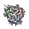 7zn0C  7zn3C 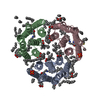 7zn9C 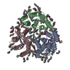 7znaC  7znbC  7zncC  7zndC  7zneC  7zngC  7znhC  7zniC  1xioS S: Starting model for refinement C: citing same article ( |
|---|---|
| Similar structure data |
-Links
 PDBj
PDBj


- Assembly
| Deposited unit | 
| |||||||||||||||||||||||||||||||||||||||||||||||||||||||||||||||||||||||||||||||||
|---|---|---|---|---|---|---|---|---|---|---|---|---|---|---|---|---|---|---|---|---|---|---|---|---|---|---|---|---|---|---|---|---|---|---|---|---|---|---|---|---|---|---|---|---|---|---|---|---|---|---|---|---|---|---|---|---|---|---|---|---|---|---|---|---|---|---|---|---|---|---|---|---|---|---|---|---|---|---|---|---|---|---|
| 1 |
| |||||||||||||||||||||||||||||||||||||||||||||||||||||||||||||||||||||||||||||||||
| Unit cell |
| |||||||||||||||||||||||||||||||||||||||||||||||||||||||||||||||||||||||||||||||||
| Noncrystallographic symmetry (NCS) | NCS domain:
NCS domain segments: Component-ID: 0 / Beg auth comp-ID: FME / Beg label comp-ID: FME / Refine code: 0
NCS ensembles :
|
-Components
-Protein , 1 types, 3 molecules ABC
| #1: Protein | Mass: 25725.627 Da / Num. of mol.: 3 Source method: isolated from a genetically manipulated source Source: (gene. exp.) Escherichia coli (E. coli) |
|---|
-Non-polymers , 6 types, 350 molecules 


| #2: Chemical | ChemComp-LFA / Icosane Mass: 282.547 Da / Num. of mol.: 50 / Source method: obtained synthetically / Formula: C20H42 Mass: 282.547 Da / Num. of mol.: 50 / Source method: obtained synthetically / Formula: C20H42#3: Chemical | ChemComp-OLA / Oleic acid Mass: 282.461 Da / Num. of mol.: 16 / Source method: obtained synthetically / Formula: C18H34O2 Mass: 282.461 Da / Num. of mol.: 16 / Source method: obtained synthetically / Formula: C18H34O2#4: Chemical | ChemComp-OLC / ( |  Mass: 356.540 Da / Num. of mol.: 1 / Source method: obtained synthetically / Formula: C21H40O4 Mass: 356.540 Da / Num. of mol.: 1 / Source method: obtained synthetically / Formula: C21H40O4#5: Chemical |  Mass: 22.990 Da / Num. of mol.: 3 / Source method: obtained synthetically / Formula: Na / Feature type: SUBJECT OF INVESTIGATION Mass: 22.990 Da / Num. of mol.: 3 / Source method: obtained synthetically / Formula: Na / Feature type: SUBJECT OF INVESTIGATION#6: Chemical | Phosphate Mass: 94.971 Da / Num. of mol.: 2 / Source method: obtained synthetically / Formula: PO4 Mass: 94.971 Da / Num. of mol.: 2 / Source method: obtained synthetically / Formula: PO4#7: Water | ChemComp-HOH / | WaterMass: 18.015 Da / Num. of mol.: 278 / Source method: isolated from a natural source / Formula: H2O |
|---|
-Details
| Has ligand of interest | Y |
|---|
-Experimental details
-Experiment
| Experiment | Method: X-RAY DIFFRACTION / Number of used crystals: 1 |
|---|
- Sample preparation
| Crystal | Density Matthews: 2.91 Å3/Da / Density % sol: 57.67 % |
|---|---|
| Crystal grow | Temperature: 293 K / Method: lipidic cubic phase Details: 0.8 M Na/K-Pi pH 8.2 crystals soaked in 1.2 M Na/K-Pi pH 7.0 before harvesting |
-Data collection
| Diffraction | Mean temperature: 100 K / Serial crystal experiment: N |
|---|---|
| Diffraction source | Source: SYNCHROTRON / Site: PETRA III, EMBL c/o DESY  / Beamline: P14 (MX2) / Wavelength: 0.976 Å / Beamline: P14 (MX2) / Wavelength: 0.976 Å |
| Detector | Type: DECTRIS EIGER X 16M / Detector: PIXEL / Date: Mar 25, 2021 |
| Radiation | Protocol: SINGLE WAVELENGTH / Monochromatic (M) / Laue (L): M / Scattering type: x-ray |
| Radiation wavelength | Wavelength: 0.976 Å / Relative weight: 1 |
| Reflection | Resolution: 2.2→49.798 Å / Num. obs: 37291 / % possible obs: 85.5 % / Redundancy: 13.1 % / CC1/2: 0.998 / Rmerge(I) obs: 0.163 / Net I/σ(I): 10.9 |
| Reflection shell | Resolution: 2.2→2.293 Å / Rmerge(I) obs: 3.775 / Mean I/σ(I) obs: 0.8 / Num. unique obs: 1865 / CC1/2: 0.5 |
- Processing
| Software |
| ||||||||||||||||||||||||||||||||||||||||||||||||||||||||||||
|---|---|---|---|---|---|---|---|---|---|---|---|---|---|---|---|---|---|---|---|---|---|---|---|---|---|---|---|---|---|---|---|---|---|---|---|---|---|---|---|---|---|---|---|---|---|---|---|---|---|---|---|---|---|---|---|---|---|---|---|---|---|
| Refinement | Method to determine structure: MOLECULAR REPLACEMENT Starting model: 1XIO Resolution: 2.2→20 Å / Cor.coef. Fo:Fc: 0.95 / Cor.coef. Fo:Fc free: 0.929 / SU B: 8.014 / SU ML: 0.188 / Cross valid method: THROUGHOUT / σ(F): 0 / ESU R: 0.384 / ESU R Free: 0.244 / Stereochemistry target values: MAXIMUM LIKELIHOOD Details: HYDROGENS HAVE BEEN ADDED IN THE RIDING POSITIONS U VALUES : REFINED INDIVIDUALLY
| ||||||||||||||||||||||||||||||||||||||||||||||||||||||||||||
| Solvent computation | Ion probe radii: 0.8 Å / Shrinkage radii: 0.8 Å / VDW probe radii: 1.2 Å / Solvent model: MASK | ||||||||||||||||||||||||||||||||||||||||||||||||||||||||||||
| Displacement parameters | Biso max: 122.27 Å2 / Biso mean: 35.505 Å2 / Biso min: 11.97 Å2
| ||||||||||||||||||||||||||||||||||||||||||||||||||||||||||||
| Refinement step | Cycle: final / Resolution: 2.2→20 Å
| ||||||||||||||||||||||||||||||||||||||||||||||||||||||||||||
| Refine LS restraints |
| ||||||||||||||||||||||||||||||||||||||||||||||||||||||||||||
| Refine LS restraints NCS | Refine-ID: X-RAY DIFFRACTION / Type: interatomic distance / Rms dev position: 0.08 Å / Weight position: 0.05
| ||||||||||||||||||||||||||||||||||||||||||||||||||||||||||||
| LS refinement shell | Resolution: 2.2→2.256 Å / Rfactor Rfree error: 0 / Total num. of bins used: 20
|
