Evidence: SAXS, Size-exclusion chromatography-coupled small-angle X-ray scattering (SEC-SAXS) was performed to assess the oligomerisation state of glycosylated (high mannose) Nariva virus receptor ...Evidence: SAXS, Size-exclusion chromatography-coupled small-angle X-ray scattering (SEC-SAXS) was performed to assess the oligomerisation state of glycosylated (high mannose) Nariva virus receptor binding protein in solution. Using the dimeric crystal structure data a best fitting model was generated using molecular dynamics, which considered the flexibility of the high mannose N-linked glycans and C- and N-termini. The extensive homodimeric interface observed in the crystal structure was largely retained.
Type
Name
Symmetry operation
Number
identity operation
1_555
x,y,z
1
Unit cell
Length a, b, c (Å)
55.382, 82.957, 92.988
Angle α, β, γ (deg.)
90.000, 90.860, 90.000
Int Tables number
4
Space group name H-M
P1211
Noncrystallographic symmetry (NCS)
NCS domain:
ID
Ens-ID
Details
1
1
chainA
2
1
(chainBandresid198through626)
NCS domain segments:
Component-ID: 1 / Ens-ID: 1 / Beg auth comp-ID: ALA / Beg label comp-ID: ALA / End auth comp-ID: ASN / End label comp-ID: ASN / Auth seq-ID: 198 - 626 / Label seq-ID: 198 - 626
Dom-ID
Selection details
Auth asym-ID
Label asym-ID
1
chainA
A
A
2
(chainBandresid198through626)
B
B
-
Components
#1: Protein
Attachmentprotein
Mass: 72484.414 Da / Num. of mol.: 2 Source method: isolated from a genetically manipulated source Source: (gene. exp.) Nariva narmovirus Cell line (production host): Human embryonic kidney 293T cells Production host: Homo sapiens (human) / References: UniProt: B8XH64
In the structure databanks used in Yorodumi, some data are registered as the other names, "COVID-19 virus" and "2019-nCoV". Here are the details of the virus and the list of structure data.
Jan 31, 2019. EMDB accession codes are about to change! (news from PDBe EMDB page)
EMDB accession codes are about to change! (news from PDBe EMDB page)
The allocation of 4 digits for EMDB accession codes will soon come to an end. Whilst these codes will remain in use, new EMDB accession codes will include an additional digit and will expand incrementally as the available range of codes is exhausted. The current 4-digit format prefixed with “EMD-” (i.e. EMD-XXXX) will advance to a 5-digit format (i.e. EMD-XXXXX), and so on. It is currently estimated that the 4-digit codes will be depleted around Spring 2019, at which point the 5-digit format will come into force.
The EM Navigator/Yorodumi systems omit the EMD- prefix.
Related info.:Q: What is EMD? / ID/Accession-code notation in Yorodumi/EM Navigator
Yorodumi is a browser for structure data from EMDB, PDB, SASBDB, etc.
This page is also the successor to EM Navigator detail page, and also detail information page/front-end page for Omokage search.
The word "yorodu" (or yorozu) is an old Japanese word meaning "ten thousand". "mi" (miru) is to see.
Related info.:EMDB / PDB / SASBDB / Comparison of 3 databanks / Yorodumi Search / Aug 31, 2016. New EM Navigator & Yorodumi / Yorodumi Papers / Jmol/JSmol / Function and homology information / Changes in new EM Navigator and Yorodumi
 Movie
Movie Controller


 Open data
 Basic information
 Components
 Keywords
 VIRAL PROTEIN /
  Function and homology information
 Nariva narmovirus
 Authors
 United Kingdom,
  Finland,
  United States, 11items
  Citation
 Structure visualization
 Downloads & links
 Other downloads
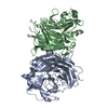
 PDBj
PDBj

 Assembly

 Mass: 18.015 Da / Num. of mol.: 721 / Source method: isolated from a natural source / Formula: H2O
 Sample preparation
 Processing