 Movie
Movie Controller


+ Open data

- Basic information
| Entry | Database: PDB / ID: 7zl9 | ||||||||||||||||||
|---|---|---|---|---|---|---|---|---|---|---|---|---|---|---|---|---|---|---|---|
| Title | Crystal structure of human GPCR Niacin receptor (HCA2) | ||||||||||||||||||
 Components Components | Hydroxycarboxylic acid receptor 2,Soluble cytochrome b562 | ||||||||||||||||||
 Keywords Keywords |  MEMBRANE PROTEIN / Niacin receptor / hydroxycarboxylic acid receptor 2 / HCA2 / GPCR / membrane protein structure MEMBRANE PROTEIN / Niacin receptor / hydroxycarboxylic acid receptor 2 / HCA2 / GPCR / membrane protein structure | ||||||||||||||||||
| Function / homology |  Function and homology informationnicotinic acid receptor activity / Hydroxycarboxylic acid-binding receptors / neutrophil apoptotic process / Class A/1 (Rhodopsin-like receptors) / positive regulation of neutrophil apoptotic process / positive regulation of adiponectin secretion / negative regulation of lipid catabolic process / cell junction / G alpha (i) signalling events / electron transfer activity ...nicotinic acid receptor activity / Hydroxycarboxylic acid-binding receptors / neutrophil apoptotic process / Class A/1 (Rhodopsin-like receptors) / positive regulation of neutrophil apoptotic process / positive regulation of adiponectin secretion / negative regulation of lipid catabolic process / cell junction / G alpha (i) signalling events / electron transfer activity / periplasmic space / iron ion binding / G protein-coupled receptor signaling pathway / heme binding / plasma membrane Function and homology informationnicotinic acid receptor activity / Hydroxycarboxylic acid-binding receptors / neutrophil apoptotic process / Class A/1 (Rhodopsin-like receptors) / positive regulation of neutrophil apoptotic process / positive regulation of adiponectin secretion / negative regulation of lipid catabolic process / cell junction / G alpha (i) signalling events / electron transfer activity ...nicotinic acid receptor activity / Hydroxycarboxylic acid-binding receptors / neutrophil apoptotic process / Class A/1 (Rhodopsin-like receptors) / positive regulation of neutrophil apoptotic process / positive regulation of adiponectin secretion / negative regulation of lipid catabolic process / cell junction / G alpha (i) signalling events / electron transfer activity / periplasmic space / iron ion binding / G protein-coupled receptor signaling pathway / heme binding / plasma membraneSimilarity search - Function | ||||||||||||||||||
| Biological species |  Homo sapiens (human) Homo sapiens (human) Escherichia coli (E. coli) Escherichia coli (E. coli) | ||||||||||||||||||
| Method | X-RAY DIFFRACTION / SYNCHROTRON / MOLECULAR REPLACEMENT / Resolution: 2.7 Å | ||||||||||||||||||
 Authors Authors | Yang, Y. / Kang, H.J. / Gao, R.G. / Wang, J.J. / Han, G.W. / FiBerto, J.F. / Wu, L.J. / Tong, J.H. / Qu, L. / Wu, Y.R. ...Yang, Y. / Kang, H.J. / Gao, R.G. / Wang, J.J. / Han, G.W. / FiBerto, J.F. / Wu, L.J. / Tong, J.H. / Qu, L. / Wu, Y.R. / Pileski, R. / Li, X.M. / Zhang, X.C. / Zhao, S.W. / Kenakin, T. / Wang, Q. / Stevens, R.C. / Peng, W. / Roth, B.L. / Rao, Z.H. / Liu, Z.J. | ||||||||||||||||||
| Funding support |  China, 5items China, 5items
| ||||||||||||||||||
 Citation Citation | Journal: Nat Commun / Year: 2023 Title: Structural insights into the human niacin receptor HCA2-G signalling complex. Authors: Yang Yang / Hye Jin Kang / Ruogu Gao / Jingjing Wang / Gye Won Han / Jeffrey F DiBerto / Lijie Wu / Jiahui Tong / Lu Qu / Yiran Wu / Ryan Pileski / Xuemei Li / Xuejun Cai Zhang / Suwen Zhao ...Authors: Yang Yang / Hye Jin Kang / Ruogu Gao / Jingjing Wang / Gye Won Han / Jeffrey F DiBerto / Lijie Wu / Jiahui Tong / Lu Qu / Yiran Wu / Ryan Pileski / Xuemei Li / Xuejun Cai Zhang / Suwen Zhao / Terry Kenakin / Quan Wang / Raymond C Stevens / Wei Peng / Bryan L Roth / Zihe Rao / Zhi-Jie Liu /   Abstract: The hydroxycarboxylic acid receptor 2 (HCA2) agonist niacin has been used as treatment for dyslipidemia for several decades albeit with skin flushing as a common side-effect in treated individuals. ...The hydroxycarboxylic acid receptor 2 (HCA2) agonist niacin has been used as treatment for dyslipidemia for several decades albeit with skin flushing as a common side-effect in treated individuals. Extensive efforts have been made to identify HCA2 targeting lipid lowering agents with fewer adverse effects, despite little being known about the molecular basis of HCA2 mediated signalling. Here, we report the cryo-electron microscopy structure of the HCA2-G signalling complex with the potent agonist MK-6892, along with crystal structures of HCA2 in inactive state. These structures, together with comprehensive pharmacological analysis, reveal the ligand binding mode and activation and signalling mechanisms of HCA2. This study elucidates the structural determinants essential for HCA2 mediated signalling and provides insights into ligand discovery for HCA2 and related receptors. | ||||||||||||||||||
| History |
|
- Structure visualization
| Structure viewer | Molecule: MolmilJmol/JSmol |
|---|
- Downloads & links
-Download
| PDBx/mmCIF format | 7zl9.cif.gz | 177.6 KB | Display | PDBx/mmCIF format |
|---|---|---|---|---|
| PDB format | pdb7zl9.ent.gz | 139.3 KB | Display | PDB format |
| PDBx/mmJSON format | 7zl9.json.gz | Tree view | PDBx/mmJSON format | |
| Others |  Other downloads Other downloads |
-Validation report
| Arichive directory | https://data.pdbj.org/pub/pdb/validation_reports/zl/7zl9ftp://data.pdbj.org/pub/pdb/validation_reports/zl/7zl9 | HTTPS FTP |
|---|
-Related structure data
| Related structure data |  7xk2C  7zlyC 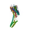 4pxzS S: Starting model for refinement C: citing same article ( |
|---|---|
| Similar structure data |

-Links
 PDBj
PDBj


- Assembly
| Deposited unit | 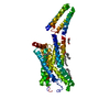
| ||||||||
|---|---|---|---|---|---|---|---|---|---|
| 1 |
| ||||||||
| Unit cell |
|
-Components
-Protein , 1 types, 1 molecules A
| #1: Protein | Mass: 49817.535 Da / Num. of mol.: 1 Source method: isolated from a genetically manipulated source Source: (gene. exp.) Homo sapiens (human), (gene. exp.) Escherichia coli (E. coli)Gene: HCAR2, GPR109A, HCA2, HM74A, NIACR1, cybC / Production host: Homo sapiens (human) / References: UniProt: Q8TDS4, UniProt: P0ABE7 |
|---|
-Non-polymers , 5 types, 15 molecules 


| #2: Chemical | ChemComp-OLA / Oleic acid Mass: 282.461 Da / Num. of mol.: 5 / Source method: obtained synthetically / Formula: C18H34O2 Mass: 282.461 Da / Num. of mol.: 5 / Source method: obtained synthetically / Formula: C18H34O2#3: Chemical | ChemComp-OLC / ( |  Mass: 356.540 Da / Num. of mol.: 1 / Source method: obtained synthetically / Formula: C21H40O4 Mass: 356.540 Da / Num. of mol.: 1 / Source method: obtained synthetically / Formula: C21H40O4#4: Chemical | ChemComp-PEG / | Diethylene glycol Mass: 106.120 Da / Num. of mol.: 1 / Source method: obtained synthetically / Formula: C4H10O3 Mass: 106.120 Da / Num. of mol.: 1 / Source method: obtained synthetically / Formula: C4H10O3#5: Chemical | ChemComp-UNX /  Num. of mol.: 5 / Source method: obtained synthetically Num. of mol.: 5 / Source method: obtained synthetically#6: Water | ChemComp-HOH / | WaterMass: 18.015 Da / Num. of mol.: 3 / Source method: isolated from a natural source / Formula: H2O |
|---|
-Details
| Has ligand of interest | N |
|---|
-Experimental details
-Experiment
| Experiment | Method: X-RAY DIFFRACTION / Number of used crystals: 1 |
|---|
- Sample preparation
| Crystal | Density Matthews: 2.85 Å3/Da / Density % sol: 56.91 % |
|---|---|
| Crystal grow | Temperature: 293 K / Method: lipidic cubic phase Details: 100mM pH5.4 sodium citrate, 60mM ammonium citrate, 36% PEG400, and 3% Additive 80 (40% PPG) |
-Data collection
| Diffraction | Mean temperature: 100 K / Serial crystal experiment: N |
|---|---|
| Diffraction source | Source: SYNCHROTRON / Site: SPring-8  / Beamline: BL41XU / Wavelength: 1 Å / Beamline: BL41XU / Wavelength: 1 Å |
| Detector | Type: RAYONIX MX225HE / Detector: CCD / Date: Jun 3, 2017 |
| Radiation | Protocol: SINGLE WAVELENGTH / Monochromatic (M) / Laue (L): M / Scattering type: x-ray |
| Radiation wavelength | Wavelength: 1 Å / Relative weight: 1 |
| Reflection | Resolution: 2.7→47.81 Å / Num. obs: 16035 / % possible obs: 100 % / Redundancy: 5.9 % / CC1/2: 0.998 / Net I/σ(I): 10.91 |
| Reflection shell | Resolution: 2.7→2.796 Å / Num. unique obs: 16035 / CC1/2: 0.998 / CC star: 1 |
- Processing
| Software |
| |||||||||||||||||||||||||||||||||||||||||||||||||||||||||||||||||||||||||||
|---|---|---|---|---|---|---|---|---|---|---|---|---|---|---|---|---|---|---|---|---|---|---|---|---|---|---|---|---|---|---|---|---|---|---|---|---|---|---|---|---|---|---|---|---|---|---|---|---|---|---|---|---|---|---|---|---|---|---|---|---|---|---|---|---|---|---|---|---|---|---|---|---|---|---|---|---|
| Refinement | Method to determine structure: MOLECULAR REPLACEMENT Starting model: 4PXZ Resolution: 2.7→30 Å / Cor.coef. Fo:Fc: 0.924 / Cor.coef. Fo:Fc free: 0.911 / Rfactor Rfree error: 0 / SU R Cruickshank DPI: 0.614 / Cross valid method: THROUGHOUT / σ(F): 0 / SU R Blow DPI: 0.601 / SU Rfree Blow DPI: 0.294 / SU Rfree Cruickshank DPI: 0.299
| |||||||||||||||||||||||||||||||||||||||||||||||||||||||||||||||||||||||||||
| Displacement parameters | Biso max: 203.34 Å2 / Biso mean: 87.66 Å2 / Biso min: 44.54 Å2
| |||||||||||||||||||||||||||||||||||||||||||||||||||||||||||||||||||||||||||
| Refine analyze | Luzzati coordinate error obs: 0.47 Å | |||||||||||||||||||||||||||||||||||||||||||||||||||||||||||||||||||||||||||
| Refinement step | Cycle: final / Resolution: 2.7→30 Å
| |||||||||||||||||||||||||||||||||||||||||||||||||||||||||||||||||||||||||||
| LS refinement shell | Resolution: 2.7→2.89 Å / Rfactor Rfree error: 0 / Total num. of bins used: 8
| |||||||||||||||||||||||||||||||||||||||||||||||||||||||||||||||||||||||||||
| Refinement TLS params. | Method: refined / Refine-ID: X-RAY DIFFRACTION
| |||||||||||||||||||||||||||||||||||||||||||||||||||||||||||||||||||||||||||
| Refinement TLS group |
|
