Journal: Nature / Year: 2022 Title: Archaic chaperone-usher pili self-secrete into superelastic zigzag springs. Authors: Natalia Pakharukova / Henri Malmi / Minna Tuittila / Tobias Dahlberg / Debnath Ghosal / Yi-Wei Chang / Si Lhyam Myint / Sari Paavilainen / Stefan David Knight / Urpo Lamminmäki / Bernt Eric ...Authors: Natalia Pakharukova / Henri Malmi / Minna Tuittila / Tobias Dahlberg / Debnath Ghosal / Yi-Wei Chang / Si Lhyam Myint / Sari Paavilainen / Stefan David Knight / Urpo Lamminmäki / Bernt Eric Uhlin / Magnus Andersson / Grant Jensen / Anton V Zavialov / Abstract: Adhesive pili assembled through the chaperone-usher pathway are hair-like appendages that mediate host tissue colonization and biofilm formation of Gram-negative bacteria. Archaic chaperone-usher ...Adhesive pili assembled through the chaperone-usher pathway are hair-like appendages that mediate host tissue colonization and biofilm formation of Gram-negative bacteria. Archaic chaperone-usher pathway pili, the most diverse and widespread chaperone-usher pathway adhesins, are promising vaccine and drug targets owing to their prevalence in the most troublesome multidrug-resistant pathogens. However, their architecture and assembly-secretion process remain unknown. Here, we present the cryo-electron microscopy structure of the prototypical archaic Csu pilus that mediates biofilm formation of Acinetobacter baumannii-a notorious multidrug-resistant nosocomial pathogen. In contrast to the thick helical tubes of the classical type 1 and P pili, archaic pili assemble into an ultrathin zigzag architecture secured by an elegant clinch mechanism. The molecular clinch provides the pilus with high mechanical stability as well as superelasticity, a property observed for the first time, to our knowledge, in biomolecules, while enabling a more economical and faster pilus production. Furthermore, we demonstrate that clinch formation at the cell surface drives pilus secretion through the outer membrane. These findings suggest that clinch-formation inhibitors might represent a new strategy to fight multidrug-resistant bacterial infections.
In the structure databanks used in Yorodumi, some data are registered as the other names, "COVID-19 virus" and "2019-nCoV". Here are the details of the virus and the list of structure data.
Jan 31, 2019. EMDB accession codes are about to change! (news from PDBe EMDB page)
EMDB accession codes are about to change! (news from PDBe EMDB page)
The allocation of 4 digits for EMDB accession codes will soon come to an end. Whilst these codes will remain in use, new EMDB accession codes will include an additional digit and will expand incrementally as the available range of codes is exhausted. The current 4-digit format prefixed with “EMD-” (i.e. EMD-XXXX) will advance to a 5-digit format (i.e. EMD-XXXXX), and so on. It is currently estimated that the 4-digit codes will be depleted around Spring 2019, at which point the 5-digit format will come into force.
The EM Navigator/Yorodumi systems omit the EMD- prefix.
Related info.:Q: What is EMD? / ID/Accession-code notation in Yorodumi/EM Navigator
Yorodumi is a browser for structure data from EMDB, PDB, SASBDB, etc.
This page is also the successor to EM Navigator detail page, and also detail information page/front-end page for Omokage search.
The word "yorodu" (or yorozu) is an old Japanese word meaning "ten thousand". "mi" (miru) is to see.
Related info.:EMDB / PDB / SASBDB / Comparison of 3 databanks / Yorodumi Search / Aug 31, 2016. New EM Navigator & Yorodumi / Yorodumi Papers / Jmol/JSmol / Function and homology information / Changes in new EM Navigator and Yorodumi
 Movie
Movie Controller

 Yorodumi
 Open data
 Basic information
 Components
 Keywords
 CELL ADHESION / chaperone-usher pathway / bacterial adhesion / biofilm formation /
  Function and homology information
 Authors
 Finland, 2items
  Citation


 Structure visualization
 Downloads & links
 Other downloads
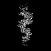

 PDBj
PDBj Assembly
 Sample preparation
 Electron microscopy imaging
 Processing