 Movie
Movie Controller

[English] 日本語
 Yorodumi
- PDB-7zky: Amyloid fibril from human systemic AA amyloidosis (vascular variant) -
+ Open data

- Basic information
| Entry | Database: PDB / ID: 7zky | ||||||
|---|---|---|---|---|---|---|---|
| Title | Amyloid fibril from human systemic AA amyloidosis (vascular variant) | ||||||
 Components Components | Amyloid protein A | ||||||
 Keywords Keywords | PROTEIN FIBRIL / Amyloids / Vascular AA / SAA / Cryo-EM | ||||||
| Function / homology |  Function and homology information Function and homology informationScavenging by Class B Receptors / positive regulation of interleukin-1 production / lymphocyte chemotaxis / high-density lipoprotein particle / Formyl peptide receptors bind formyl peptides and many other ligands / macrophage chemotaxis / regulation of protein secretion / TRAF6 mediated NF-kB activation / Advanced glycosylation endproduct receptor signaling / positive regulation of cell adhesion ...Scavenging by Class B Receptors / positive regulation of interleukin-1 production / lymphocyte chemotaxis / high-density lipoprotein particle / Formyl peptide receptors bind formyl peptides and many other ligands / macrophage chemotaxis / regulation of protein secretion / TRAF6 mediated NF-kB activation / Advanced glycosylation endproduct receptor signaling / positive regulation of cell adhesion / cytoplasmic microtubule / endocytic vesicle lumen / neutrophil chemotaxis / G protein-coupled receptor binding / positive regulation of cytokine production / acute-phase response / TAK1-dependent IKK and NF-kappa-B activation / negative regulation of inflammatory response / platelet activation / heparin binding / positive regulation of cytosolic calcium ion concentration / G alpha (i) signalling events / G alpha (q) signalling events / Interleukin-4 and Interleukin-13 signaling / Amyloid fiber formation / extracellular exosome / extracellular regionSimilarity search - Function | ||||||
| Biological species |  Homo sapiens (human) Homo sapiens (human) | ||||||
| Method | ELECTRON MICROSCOPY / helical reconstruction / cryo EM / Resolution: 2.56 Å | ||||||
 Authors Authors | Banerjee, S. / Schmidt, M. / Faendrich, M. | ||||||
| Funding support | 1items
| ||||||
 Citation Citation | Journal: Nat Commun / Year: 2022 Title: Amyloid fibril structure from the vascular variant of systemic AA amyloidosis. Authors: Sambhasan Banerjee / Julian Baur / Christoph Daniel / Peter Benedikt Pfeiffer / Manuel Hitzenberger / Lukas Kuhn / Sebastian Wiese / Johan Bijzet / Christian Haupt / Kerstin U Amann / Martin ...Authors: Sambhasan Banerjee / Julian Baur / Christoph Daniel / Peter Benedikt Pfeiffer / Manuel Hitzenberger / Lukas Kuhn / Sebastian Wiese / Johan Bijzet / Christian Haupt / Kerstin U Amann / Martin Zacharias / Bouke P C Hazenberg / Gunilla T Westermark / Matthias Schmidt / Marcus Fändrich /    Abstract: Systemic AA amyloidosis is a debilitating protein misfolding disease in humans and animals. In humans, it occurs in two variants that are called 'vascular' and 'glomerular', depending on the main ...Systemic AA amyloidosis is a debilitating protein misfolding disease in humans and animals. In humans, it occurs in two variants that are called 'vascular' and 'glomerular', depending on the main amyloid deposition site in the kidneys. Using cryo electron microscopy, we here show the amyloid fibril structure underlying the vascular disease variant. Fibrils purified from the tissue of such patients are mainly left-hand twisted and contain two non-equal stacks of fibril proteins. They contrast in these properties to the fibrils from the glomerular disease variant which are right-hand twisted and consist of two structurally equal stacks of fibril proteins. Our data demonstrate that the different disease variants in systemic AA amyloidosis are associated with different fibril morphologies. | ||||||
| History |
|
- Structure visualization
| Structure viewer | Molecule: MolmilJmol/JSmol |
|---|
- Downloads & links
-Download
| PDBx/mmCIF format | 7zky.cif.gz | 116.2 KB | Display | PDBx/mmCIF format |
|---|---|---|---|---|
| PDB format | pdb7zky.ent.gz | 96.5 KB | Display | PDB format |
| PDBx/mmJSON format | 7zky.json.gz | Tree view | PDBx/mmJSON format | |
| Others |  Other downloads Other downloads |
-Validation report
| Arichive directory | https://data.pdbj.org/pub/pdb/validation_reports/zk/7zkyftp://data.pdbj.org/pub/pdb/validation_reports/zk/7zky | HTTPS FTP |
|---|
-Related structure data
| Related structure data | 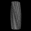 14771MC M: map data used to model this data C: citing same article ( |
|---|---|
| Similar structure data |
-Links
 PDBj
PDBj


- Assembly
| Deposited unit | 
|
|---|---|
| 1 |
|
-Components
| #1: Protein | / Amyloid fibril protein AA Mass: 7775.502 Da / Num. of mol.: 12 / Source method: isolated from a natural source / Source: (natural) Homo sapiens (human) / References: UniProt: P0DJI8 |
|---|
-Experimental details
-Experiment
| Experiment | Method: ELECTRON MICROSCOPY |
|---|---|
| EM experiment | Aggregation state: HELICAL ARRAY / 3D reconstruction method: helical reconstruction |
- Sample preparation
| Component | Name: Amyloid fibril from human systemic AA amyloidosis (vascular variant)Amyloid Type: COMPLEX / Entity ID: all / Source: NATURAL |
|---|---|
| Source (natural) | Organism: Homo sapiens (human) |
| Buffer solution | pH: 7 |
| Specimen | Embedding applied: NO / Shadowing applied: NO / Staining applied: NO / Vitrification applied: YES |
| Specimen support | Grid material: COPPER / Grid mesh size: 400 divisions/in. / Grid type: C-flat-1.2/1.3 |
| Vitrification | Cryogen name: ETHANE |
- Electron microscopy imaging
| Experimental equipment |  Model: Titan Krios / Image courtesy: FEI Company |
|---|---|
| Microscopy | Model: FEI TITAN KRIOS |
| Electron gun | Electron source: FIELD EMISSION GUN / Accelerating voltage: 300 kV / Illumination mode: SPOT SCAN |
| Electron lens | Mode: BRIGHT FIELDBright-field microscopy / Nominal magnification: 130000 X / Nominal defocus max: 2000 nm / Nominal defocus min: 800 nm / Cs: 2.7 mm |
| Image recording | Average exposure time: 10 sec. / Electron dose: 42.84 e/Å2 / Detector mode: COUNTING / Film or detector model: GATAN K2 SUMMIT (4k x 4k) |
- Processing
| CTF correction | Type: PHASE FLIPPING AND AMPLITUDE CORRECTION |
|---|---|
| Helical symmerty | Angular rotation/subunit: -1.63 ° / Axial rise/subunit: 4.75 Å / Axial symmetry: C1 |
| 3D reconstruction | Resolution: 2.56 Å / Resolution method: FSC 0.143 CUT-OFF / Num. of particles: 77061 / Algorithm: SIMULTANEOUS ITERATIVE (SIRT) / Num. of class averages: 1 / Symmetry type: HELICAL |
| Atomic model building | Protocol: AB INITIO MODEL |
