 Movie
Movie Controller

[English] 日本語
 Yorodumi
- PDB-7zkt: Moss spermine/spermidine acetyl transferase (PpSSAT) in complex w... -
+ Open data

- Basic information
| Entry | Database: PDB / ID: 7zkt | ||||||
|---|---|---|---|---|---|---|---|
| Title | Moss spermine/spermidine acetyl transferase (PpSSAT) in complex with CoA and lysine | ||||||
 Components Components | N-acetyltransferase domain-containing protein | ||||||
 Keywords Keywords |  TRANSFERASE / Acetylation / moss / SSAT TRANSFERASE / Acetylation / moss / SSAT | ||||||
| Function / homology | N-acetyltransferase activity / Acetyltransferase (GNAT) family / Gcn5-related N-acetyltransferase (GNAT) domain profile. / GNAT domain / Acyl-CoA N-acyltransferase / COENZYME A / LYSINE / N-acetyltransferase domain-containing protein Function and homology information Function and homology information | ||||||
| Biological species |  Physcomitrium patens (plant) Physcomitrium patens (plant) | ||||||
| Method | X-RAY DIFFRACTION / SYNCHROTRON / MOLECULAR REPLACEMENT / molecular replacement / Resolution: 2.06 Å | ||||||
 Authors Authors | Morera, S. / Kopecny, D. / Vigouroux, A. / Briozzo, P. | ||||||
| Funding support |  France, 1items France, 1items
| ||||||
 Citation Citation | Journal: Plant J. / Year: 2023 Title: Biochemical and structural basis of polyamine, lysine and ornithine acetylation catalyzed by spermine/spermidine N-acetyl transferase in moss and maize. Authors: Belicek, J. / Luptakova, E. / Kopecny, D. / Frommel, J. / Vigouroux, A. / Cavar Zeljkovic, S. / Jagic, F. / Briozzo, P. / Kopecny, D.J. / Tarkowski, P. / Nisler, J. / De Diego, N. / Morera, S. / Kopecna, M. | ||||||
| History |
|
- Structure visualization
| Structure viewer | Molecule: MolmilJmol/JSmol |
|---|
- Downloads & links
-Download
| PDBx/mmCIF format | 7zkt.cif.gz | 686.8 KB | Display | PDBx/mmCIF format |
|---|---|---|---|---|
| PDB format | pdb7zkt.ent.gz | 576.1 KB | Display | PDB format |
| PDBx/mmJSON format | 7zkt.json.gz | Tree view | PDBx/mmJSON format | |
| Others |  Other downloads Other downloads |
-Validation report
| Arichive directory | https://data.pdbj.org/pub/pdb/validation_reports/zk/7zktftp://data.pdbj.org/pub/pdb/validation_reports/zk/7zkt | HTTPS FTP |
|---|
-Related structure data
| Related structure data | 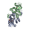 7zhcSC S: Starting model for refinement C: citing same article ( |
|---|---|
| Similar structure data |
-Links
 PDBj
PDBj


- Assembly
| Deposited unit | 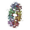
| ||||||||
|---|---|---|---|---|---|---|---|---|---|
| 1 | 
| ||||||||
| 2 | 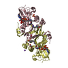
| ||||||||
| 3 | 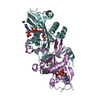
| ||||||||
| 4 | 
| ||||||||
| Unit cell |
|
-Components
-Protein , 1 types, 8 molecules ABCDEFGH
| #1: Protein | Mass: 26103.910 Da / Num. of mol.: 8 Source method: isolated from a genetically manipulated source Source: (gene. exp.) Physcomitrium patens (plant) / Gene: PHYPA_008003 / Production host:  Escherichia coli BL21(DE3) (bacteria) / References: UniProt: A0A2K1KKM6 Escherichia coli BL21(DE3) (bacteria) / References: UniProt: A0A2K1KKM6 |
|---|
-Non-polymers , 6 types, 716 molecules 


| #2: Chemical | ChemComp-COA / Coenzyme A Mass: 767.534 Da / Num. of mol.: 8 / Source method: obtained synthetically / Formula: C21H36N7O16P3S / Feature type: SUBJECT OF INVESTIGATION Mass: 767.534 Da / Num. of mol.: 8 / Source method: obtained synthetically / Formula: C21H36N7O16P3S / Feature type: SUBJECT OF INVESTIGATION#3: Chemical | ChemComp-LYS / Lysine Type: L-peptide linking / Mass: 147.195 Da / Num. of mol.: 8 / Source method: obtained synthetically / Formula: C6H15N2O2 / Feature type: SUBJECT OF INVESTIGATION Type: L-peptide linking / Mass: 147.195 Da / Num. of mol.: 8 / Source method: obtained synthetically / Formula: C6H15N2O2 / Feature type: SUBJECT OF INVESTIGATION#4: Chemical | ChemComp-NA /  Mass: 22.990 Da / Num. of mol.: 9 / Source method: obtained synthetically / Formula: Na Mass: 22.990 Da / Num. of mol.: 9 / Source method: obtained synthetically / Formula: Na#5: Chemical | ChemComp-EDO / Ethylene glycol Mass: 62.068 Da / Num. of mol.: 7 / Source method: obtained synthetically / Formula: C2H6O2 Mass: 62.068 Da / Num. of mol.: 7 / Source method: obtained synthetically / Formula: C2H6O2#6: Chemical | ChemComp-SO4 / Sulfate Mass: 96.063 Da / Num. of mol.: 6 / Source method: obtained synthetically / Formula: SO4 Mass: 96.063 Da / Num. of mol.: 6 / Source method: obtained synthetically / Formula: SO4#7: Water | ChemComp-HOH / | WaterMass: 18.015 Da / Num. of mol.: 678 / Source method: isolated from a natural source / Formula: H2O |
|---|
-Details
| Has ligand of interest | Y |
|---|
-Experimental details
-Experiment
| Experiment | Method: X-RAY DIFFRACTION / Number of used crystals: 1 |
|---|
- Sample preparation
| Crystal | Density Matthews: 2.79 Å3/Da / Density % sol: 55.91 % |
|---|---|
| Crystal grow | Temperature: 292 K / Method: vapor diffusion, hanging drop Details: 0.2 M ammonium sulfate, 0.1 M trisodium citrate pH 5.6, 15 % PEG 4000 |
-Data collection
| Diffraction | Mean temperature: 100 K / Serial crystal experiment: N |
|---|---|
| Diffraction source | Source: SYNCHROTRON / Site: SOLEIL / Beamline: PROXIMA 2 / Wavelength: 0.98011 Å |
| Detector | Type: DECTRIS EIGER X 16M / Detector: PIXEL / Date: Mar 18, 2022 |
| Radiation | Protocol: SINGLE WAVELENGTH / Monochromatic (M) / Laue (L): M / Scattering type: x-ray |
| Radiation wavelength | Wavelength: 0.98011 Å / Relative weight: 1 |
| Reflection | Resolution: 2.06→94.55 Å / Num. obs: 138210 / % possible obs: 98.2 % / Redundancy: 6.2 % / Biso Wilson estimate: 44.92 Å2 / CC1/2: 0.996 / Rmerge(I) obs: 0.161 / Net I/σ(I): 7.1 |
| Reflection shell | Resolution: 2.06→2.291 Å / Rmerge(I) obs: 1.2 / Num. unique obs: 4684 / CC1/2: 0.579 |
-Phasing
| Phasing | Method: molecular replacement |
|---|
- Processing
| Software |
| |||||||||||||||||||||||||||||||||||||||||||||||||||||||||||||||||||||||||||||||||||||||||||||||||||||||||||||||||||||||||||||||||||||||||||||||||||||||||||||||||||||||||||||||||||||||||||||||||||||||||||||||||||||||||||||||||
|---|---|---|---|---|---|---|---|---|---|---|---|---|---|---|---|---|---|---|---|---|---|---|---|---|---|---|---|---|---|---|---|---|---|---|---|---|---|---|---|---|---|---|---|---|---|---|---|---|---|---|---|---|---|---|---|---|---|---|---|---|---|---|---|---|---|---|---|---|---|---|---|---|---|---|---|---|---|---|---|---|---|---|---|---|---|---|---|---|---|---|---|---|---|---|---|---|---|---|---|---|---|---|---|---|---|---|---|---|---|---|---|---|---|---|---|---|---|---|---|---|---|---|---|---|---|---|---|---|---|---|---|---|---|---|---|---|---|---|---|---|---|---|---|---|---|---|---|---|---|---|---|---|---|---|---|---|---|---|---|---|---|---|---|---|---|---|---|---|---|---|---|---|---|---|---|---|---|---|---|---|---|---|---|---|---|---|---|---|---|---|---|---|---|---|---|---|---|---|---|---|---|---|---|---|---|---|---|---|---|---|---|---|---|---|---|---|---|---|---|---|---|---|---|---|---|---|
| Refinement | Method to determine structure: MOLECULAR REPLACEMENT Starting model: 7ZHC Resolution: 2.06→94.55 Å / Cor.coef. Fo:Fc: 0.951 / Cor.coef. Fo:Fc free: 0.941 / SU R Cruickshank DPI: 0.258 / Cross valid method: THROUGHOUT / σ(F): 0 / SU R Blow DPI: 0.256 / SU Rfree Blow DPI: 0.183 / SU Rfree Cruickshank DPI: 0.185
| |||||||||||||||||||||||||||||||||||||||||||||||||||||||||||||||||||||||||||||||||||||||||||||||||||||||||||||||||||||||||||||||||||||||||||||||||||||||||||||||||||||||||||||||||||||||||||||||||||||||||||||||||||||||||||||||||
| Displacement parameters | Biso max: 188.9 Å2 / Biso mean: 55.78 Å2 / Biso min: 19.69 Å2
| |||||||||||||||||||||||||||||||||||||||||||||||||||||||||||||||||||||||||||||||||||||||||||||||||||||||||||||||||||||||||||||||||||||||||||||||||||||||||||||||||||||||||||||||||||||||||||||||||||||||||||||||||||||||||||||||||
| Refine analyze | Luzzati coordinate error obs: 0.26 Å | |||||||||||||||||||||||||||||||||||||||||||||||||||||||||||||||||||||||||||||||||||||||||||||||||||||||||||||||||||||||||||||||||||||||||||||||||||||||||||||||||||||||||||||||||||||||||||||||||||||||||||||||||||||||||||||||||
| Refinement step | Cycle: final / Resolution: 2.06→94.55 Å
| |||||||||||||||||||||||||||||||||||||||||||||||||||||||||||||||||||||||||||||||||||||||||||||||||||||||||||||||||||||||||||||||||||||||||||||||||||||||||||||||||||||||||||||||||||||||||||||||||||||||||||||||||||||||||||||||||
| Refine LS restraints |
| |||||||||||||||||||||||||||||||||||||||||||||||||||||||||||||||||||||||||||||||||||||||||||||||||||||||||||||||||||||||||||||||||||||||||||||||||||||||||||||||||||||||||||||||||||||||||||||||||||||||||||||||||||||||||||||||||
| LS refinement shell | Resolution: 2.06→2.12 Å / Rfactor Rfree error: 0 / Total num. of bins used: 20
| |||||||||||||||||||||||||||||||||||||||||||||||||||||||||||||||||||||||||||||||||||||||||||||||||||||||||||||||||||||||||||||||||||||||||||||||||||||||||||||||||||||||||||||||||||||||||||||||||||||||||||||||||||||||||||||||||
| Refinement TLS params. | Method: refined / Refine-ID: X-RAY DIFFRACTION
| |||||||||||||||||||||||||||||||||||||||||||||||||||||||||||||||||||||||||||||||||||||||||||||||||||||||||||||||||||||||||||||||||||||||||||||||||||||||||||||||||||||||||||||||||||||||||||||||||||||||||||||||||||||||||||||||||
| Refinement TLS group |
|
