 Movie
Movie Controller

[English] 日本語
 Yorodumi
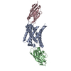
- PDB-7zk1: Crystal structure of cystinosin from Arabidopsis thaliana bound t... -
+ Open data

- Basic information
| Entry | Database: PDB / ID: 7zk1 | ||||||
|---|---|---|---|---|---|---|---|
| Title | Crystal structure of cystinosin from Arabidopsis thaliana bound to sybody and nanobody | ||||||
 Components Components |
| ||||||
 Keywords Keywords |  MEMBRANE PROTEIN / Cystinosin / PQ-loop protein / proton coupling / cystine transport MEMBRANE PROTEIN / Cystinosin / PQ-loop protein / proton coupling / cystine transport | ||||||
| Function / homology | Lysosomal cystine transporter / Repeated motif present between transmembrane helices in cystinosin, yeast ERS1p, mannose-P-dolichol utilization defect 1, and other hypothetical proteins. / PQ-loop repeat / PQ loop repeat / plant-type vacuole / lysosomal membrane / Cystinosin homolog Function and homology information Function and homology information | ||||||
| Biological species |  Arabidopsis thaliana (thale cress) Arabidopsis thaliana (thale cress) Lama glama (llama) Lama glama (llama)synthetic construct (others) | ||||||
| Method | X-RAY DIFFRACTION / SYNCHROTRON / SAD / Resolution: 2.65 Å | ||||||
 Authors Authors | Loebel, M. / Newstead, S. / Omari, K.E. | ||||||
| Funding support |  United Kingdom, 1items United Kingdom, 1items
| ||||||
 Citation Citation | Journal: Nat Commun / Year: 2022 Title: Structural basis for proton coupled cystine transport by cystinosin. Authors: Lobel, M. / Salphati, S.P. / El Omari, K. / Wagner, A. / Tucker, S.J. / Parker, J.L. / Newstead, S. | ||||||
| History |
|
- Structure visualization
| Structure viewer | Molecule: MolmilJmol/JSmol |
|---|
- Downloads & links
-Download
| PDBx/mmCIF format | 7zk1.cif.gz | 205 KB | Display | PDBx/mmCIF format |
|---|---|---|---|---|
| PDB format | pdb7zk1.ent.gz | 168.4 KB | Display | PDB format |
| PDBx/mmJSON format | 7zk1.json.gz | Tree view | PDBx/mmJSON format | |
| Others |  Other downloads Other downloads |
-Validation report
| Arichive directory | https://data.pdbj.org/pub/pdb/validation_reports/zk/7zk1ftp://data.pdbj.org/pub/pdb/validation_reports/zk/7zk1 | HTTPS FTP |
|---|
-Related structure data

-Links
 PDBj
PDBj

- Assembly
| Deposited unit | 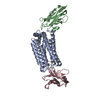
| ||||||||
|---|---|---|---|---|---|---|---|---|---|
| 1 |
| ||||||||
| Unit cell |
|
-Components
| #1: Protein | Mass: 31902.965 Da / Num. of mol.: 1 Source method: isolated from a genetically manipulated source Source: (gene. exp.) Arabidopsis thaliana (thale cress) / Gene: At5g40670, MNF13.23 / Production host:  Saccharomyces cerevisiae (brewer's yeast) / Strain (production host): BJ5460 / References: UniProt: P57758 Saccharomyces cerevisiae (brewer's yeast) / Strain (production host): BJ5460 / References: UniProt: P57758 |
|---|---|
| #2: Antibody | Mass: 13888.493 Da / Num. of mol.: 1 Source method: isolated from a genetically manipulated source Source: (gene. exp.) Lama glama (llama) / Production host:  Escherichia coli MC1061 (bacteria) Escherichia coli MC1061 (bacteria) |
| #3: Antibody | Mass: 13651.062 Da / Num. of mol.: 1 Source method: isolated from a genetically manipulated source Source: (gene. exp.) synthetic construct (others) / Production host: Escherichia coli (E. coli) |
| #4: Water | ChemComp-HOH / Water Mass: 18.015 Da / Num. of mol.: 32 / Source method: isolated from a natural source / Formula: H2O Mass: 18.015 Da / Num. of mol.: 32 / Source method: isolated from a natural source / Formula: H2O |
-Experimental details
-Experiment
| Experiment | Method: X-RAY DIFFRACTION / Number of used crystals: 1 |
|---|
- Sample preparation
| Crystal | Density Matthews: 4.13 Å3/Da / Density % sol: 70.19 % |
|---|---|
| Crystal grow | Temperature: 293 K / Method: lipidic cubic phase / pH: 5.5 Details: 27.5% PEG 500DME, 100 mM MES-NaOH, pH 5.50, 100 mM K-formate |
-Data collection
| Diffraction | Mean temperature: 100 K / Serial crystal experiment: N | |||||||||||||||||||||||||||
|---|---|---|---|---|---|---|---|---|---|---|---|---|---|---|---|---|---|---|---|---|---|---|---|---|---|---|---|---|
| Diffraction source | Source: SYNCHROTRON / Site: Diamond / Beamline: I24 / Wavelength: 0.9999 Å | |||||||||||||||||||||||||||
| Detector | Type: DECTRIS PILATUS3 6M / Detector: PIXEL / Date: Jan 20, 2021 | |||||||||||||||||||||||||||
| Radiation | Protocol: SINGLE WAVELENGTH / Monochromatic (M) / Laue (L): M / Scattering type: x-ray | |||||||||||||||||||||||||||
| Radiation wavelength | Wavelength: 0.9999 Å / Relative weight: 1 | |||||||||||||||||||||||||||
| Reflection | Resolution: 2.65→79.99 Å / Num. obs: 28079 / % possible obs: 99.9 % / Redundancy: 20 % / CC1/2: 0.996 / Rmerge(I) obs: 0.542 / Rpim(I) all: 0.114 / Rrim(I) all: 0.554 / Net I/σ(I): 5.9 | |||||||||||||||||||||||||||
| Reflection shell | Diffraction-ID: 1 / % possible all: 99.9
|
- Processing
| Software |
| ||||||||||||||||||||||||||||||||||||||||||||||||||||||||||||||||||||||||||||||||||||||||||||||||||||
|---|---|---|---|---|---|---|---|---|---|---|---|---|---|---|---|---|---|---|---|---|---|---|---|---|---|---|---|---|---|---|---|---|---|---|---|---|---|---|---|---|---|---|---|---|---|---|---|---|---|---|---|---|---|---|---|---|---|---|---|---|---|---|---|---|---|---|---|---|---|---|---|---|---|---|---|---|---|---|---|---|---|---|---|---|---|---|---|---|---|---|---|---|---|---|---|---|---|---|---|---|---|
| Refinement | Method to determine structure: SAD / Resolution: 2.65→79.99 Å / SU R Cruickshank DPI: 0.391 / Cross valid method: THROUGHOUT / SU R Blow DPI: 0.391 / SU Rfree Blow DPI: 0.286 / SU Rfree Cruickshank DPI: 0.289
| ||||||||||||||||||||||||||||||||||||||||||||||||||||||||||||||||||||||||||||||||||||||||||||||||||||
| Displacement parameters | Biso max: 131.86 Å2 / Biso mean: 84.8151 Å2 / Biso min: 33.68 Å2
| ||||||||||||||||||||||||||||||||||||||||||||||||||||||||||||||||||||||||||||||||||||||||||||||||||||
| Refine analyze | Luzzati coordinate error obs: 0.49 Å | ||||||||||||||||||||||||||||||||||||||||||||||||||||||||||||||||||||||||||||||||||||||||||||||||||||
| Refinement step | Cycle: LAST / Resolution: 2.65→79.99 Å
| ||||||||||||||||||||||||||||||||||||||||||||||||||||||||||||||||||||||||||||||||||||||||||||||||||||
| LS refinement shell | Resolution: 2.65→2.68 Å
| ||||||||||||||||||||||||||||||||||||||||||||||||||||||||||||||||||||||||||||||||||||||||||||||||||||
| Refinement TLS params. | Method: refined / Refine-ID: X-RAY DIFFRACTION
| ||||||||||||||||||||||||||||||||||||||||||||||||||||||||||||||||||||||||||||||||||||||||||||||||||||
| Refinement TLS group |
|
