 Movie
Movie Controller

[English] 日本語
 Yorodumi
- PDB-7zia: Crystal structure of dCK C4S-S74E mutant in complex with UDP and ... -
+ Open data

- Basic information
| Entry | Database: PDB / ID: 7zia | ||||||
|---|---|---|---|---|---|---|---|
| Title | Crystal structure of dCK C4S-S74E mutant in complex with UDP and the OR0634 inhibitor | ||||||
 Components Components | Deoxycytidine kinase | ||||||
 Keywords Keywords | TRANSFERASE / Inhibitor | ||||||
| Function / homology |  Function and homology informationdeoxycytidine kinase / 2'-deoxyadenosine kinase / deoxyguanosine kinase / dAMP salvage / deoxycytidine kinase activity / nucleoside phosphate biosynthetic process / deoxyguanosine kinase activity / deoxyadenosine kinase activity / Pyrimidine salvage / cytidine kinase activity ...deoxycytidine kinase / 2'-deoxyadenosine kinase / deoxyguanosine kinase / dAMP salvage / deoxycytidine kinase activity / nucleoside phosphate biosynthetic process / deoxyguanosine kinase activity / deoxyadenosine kinase activity / Pyrimidine salvage / cytidine kinase activity / pyrimidine nucleotide metabolic process / Purine salvage / phosphorylation / protein homodimerization activity / mitochondrion / nucleoplasm / ATP binding / cytosol / cytoplasm Function and homology informationdeoxycytidine kinase / 2'-deoxyadenosine kinase / deoxyguanosine kinase / dAMP salvage / deoxycytidine kinase activity / nucleoside phosphate biosynthetic process / deoxyguanosine kinase activity / deoxyadenosine kinase activity / Pyrimidine salvage / cytidine kinase activity ...deoxycytidine kinase / 2'-deoxyadenosine kinase / deoxyguanosine kinase / dAMP salvage / deoxycytidine kinase activity / nucleoside phosphate biosynthetic process / deoxyguanosine kinase activity / deoxyadenosine kinase activity / Pyrimidine salvage / cytidine kinase activity / pyrimidine nucleotide metabolic process / Purine salvage / phosphorylation / protein homodimerization activity / mitochondrion / nucleoplasm / ATP binding / cytosol / cytoplasmSimilarity search - Function | ||||||
| Biological species |  Homo sapiens (human) Homo sapiens (human) | ||||||
| Method | X-RAY DIFFRACTION / SYNCHROTRON / MOLECULAR REPLACEMENT / Resolution: 1.7 Å | ||||||
 Authors Authors | Ben-Yaala, K. / Saez-Ayala, M. / Betzi, S. / Rebuffet, E. / Morelli, X. | ||||||
| Funding support |  France, 1items France, 1items
| ||||||
 Citation Citation | Journal: Nat Commun / Year: 2023 Title: From a drug repositioning to a structure-based drug design approach to tackle acute lymphoblastic leukemia. Authors: Saez-Ayala, M. / Hoffer, L. / Abel, S. / Ben Yaala, K. / Sicard, B. / Andrieu, G.P. / Latiri, M. / Davison, E.K. / Ciufolini, M.A. / Bremond, P. / Rebuffet, E. / Roche, P. / Derviaux, C. / ...Authors: Saez-Ayala, M. / Hoffer, L. / Abel, S. / Ben Yaala, K. / Sicard, B. / Andrieu, G.P. / Latiri, M. / Davison, E.K. / Ciufolini, M.A. / Bremond, P. / Rebuffet, E. / Roche, P. / Derviaux, C. / Voisset, E. / Montersino, C. / Castellano, R. / Collette, Y. / Asnafi, V. / Betzi, S. / Dubreuil, P. / Combes, S. / Morelli, X. | ||||||
| History |
|
- Structure visualization
| Structure viewer | Molecule: MolmilJmol/JSmol |
|---|
- Downloads & links
-Download
| PDBx/mmCIF format | 7zia.cif.gz | 70.5 KB | Display | PDBx/mmCIF format |
|---|---|---|---|---|
| PDB format | pdb7zia.ent.gz | 48.6 KB | Display | PDB format |
| PDBx/mmJSON format | 7zia.json.gz | Tree view | PDBx/mmJSON format | |
| Others |  Other downloads Other downloads |
-Validation report
| Arichive directory | https://data.pdbj.org/pub/pdb/validation_reports/zi/7ziaftp://data.pdbj.org/pub/pdb/validation_reports/zi/7zia | HTTPS FTP |
|---|
-Related structure data
| Related structure data | 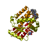 7zi1C 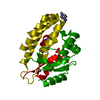 7zi2C  7zi3C 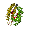 7zi5C 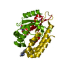 7zi6C 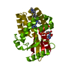 7zi7C  7zi8C  7zi9C  7zibC 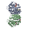 4kcgS S: Starting model for refinement C: citing same article ( |
|---|---|
| Similar structure data |
-Links
 PDBj
PDBj


- Assembly
| Deposited unit | 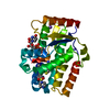
| ||||||||
|---|---|---|---|---|---|---|---|---|---|
| 1 | 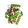
| ||||||||
| Unit cell |
|
-Components
| #1: Protein | / dCK / Deoxyadenosine kinase / Deoxyguanosine kinase Mass: 32701.627 Da / Num. of mol.: 1 Source method: isolated from a genetically manipulated source Source: (gene. exp.) Homo sapiens (human) / Gene: DCK / Production host:  Escherichia coli (E. coli) Escherichia coli (E. coli)References: UniProt: P27707, deoxycytidine kinase, 2'-deoxyadenosine kinase, deoxyguanosine kinase |
|---|---|
| #2: Chemical | ChemComp-UDP / Uridine diphosphate  Type: RNA linking / Mass: 404.161 Da / Num. of mol.: 1 / Source method: obtained synthetically / Formula: C9H14N2O12P2 / Comment: UDP*YM Type: RNA linking / Mass: 404.161 Da / Num. of mol.: 1 / Source method: obtained synthetically / Formula: C9H14N2O12P2 / Comment: UDP*YM |
| #3: Chemical | ChemComp-J29 / 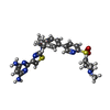  Mass: 579.740 Da / Num. of mol.: 1 / Source method: obtained synthetically / Formula: C27H33N9O2S2 / Feature type: SUBJECT OF INVESTIGATION Mass: 579.740 Da / Num. of mol.: 1 / Source method: obtained synthetically / Formula: C27H33N9O2S2 / Feature type: SUBJECT OF INVESTIGATION |
| #4: Chemical | ChemComp-NA /   Mass: 22.990 Da / Num. of mol.: 1 / Source method: obtained synthetically / Formula: Na Mass: 22.990 Da / Num. of mol.: 1 / Source method: obtained synthetically / Formula: Na |
| #5: Water | ChemComp-HOH / Water Mass: 18.015 Da / Num. of mol.: 99 / Source method: isolated from a natural source / Formula: H2O Mass: 18.015 Da / Num. of mol.: 99 / Source method: isolated from a natural source / Formula: H2O |
| Has ligand of interest | Y |
-Experimental details
-Experiment
| Experiment | Method: X-RAY DIFFRACTION / Number of used crystals: 1 |
|---|
- Sample preparation
| Crystal | Density Matthews: 2.21 Å3/Da / Density % sol: 44.22 % |
|---|---|
| Crystal grow | Temperature: 285 K / Method: vapor diffusion, hanging drop / pH: 7.5 / Details: 0.9 M Sodium citrate, 60 mM HEPES |
-Data collection
| Diffraction | Mean temperature: 100 K / Serial crystal experiment: N | ||||||||||||||||||||||||||||||||||||||||||||||||||||||||||||||||||||||||||||||||||||||||||||||||||||
|---|---|---|---|---|---|---|---|---|---|---|---|---|---|---|---|---|---|---|---|---|---|---|---|---|---|---|---|---|---|---|---|---|---|---|---|---|---|---|---|---|---|---|---|---|---|---|---|---|---|---|---|---|---|---|---|---|---|---|---|---|---|---|---|---|---|---|---|---|---|---|---|---|---|---|---|---|---|---|---|---|---|---|---|---|---|---|---|---|---|---|---|---|---|---|---|---|---|---|---|---|---|
| Diffraction source | Source: SYNCHROTRON / Site: SOLEIL / Beamline: PROXIMA 2 / Wavelength: 0.98013 Å | ||||||||||||||||||||||||||||||||||||||||||||||||||||||||||||||||||||||||||||||||||||||||||||||||||||
| Detector | Type: DECTRIS EIGER X 9M / Detector: PIXEL / Date: Nov 7, 2020 | ||||||||||||||||||||||||||||||||||||||||||||||||||||||||||||||||||||||||||||||||||||||||||||||||||||
| Radiation | Protocol: SINGLE WAVELENGTH / Monochromatic (M) / Laue (L): M / Scattering type: x-ray | ||||||||||||||||||||||||||||||||||||||||||||||||||||||||||||||||||||||||||||||||||||||||||||||||||||
| Radiation wavelength | Wavelength: 0.98013 Å / Relative weight: 1 | ||||||||||||||||||||||||||||||||||||||||||||||||||||||||||||||||||||||||||||||||||||||||||||||||||||
| Reflection | Resolution: 1.7→48.79 Å / Num. obs: 32937 / % possible obs: 99.8 % / Redundancy: 25.053 % / Biso Wilson estimate: 44.045 Å2 / CC1/2: 1 / Rmerge(I) obs: 0.057 / Rrim(I) all: 0.058 / Χ2: 0.822 / Net I/σ(I): 28.58 / Num. measured all: 825173 | ||||||||||||||||||||||||||||||||||||||||||||||||||||||||||||||||||||||||||||||||||||||||||||||||||||
| Reflection shell | Diffraction-ID: 1
|
- Processing
| Software |
| ||||||||||||||||||||||||||||||||||||||||||||||||||||||||||||
|---|---|---|---|---|---|---|---|---|---|---|---|---|---|---|---|---|---|---|---|---|---|---|---|---|---|---|---|---|---|---|---|---|---|---|---|---|---|---|---|---|---|---|---|---|---|---|---|---|---|---|---|---|---|---|---|---|---|---|---|---|---|
| Refinement | Method to determine structure: MOLECULAR REPLACEMENT Starting model: 4KCG Resolution: 1.7→48.79 Å / Cor.coef. Fo:Fc: 0.972 / Cor.coef. Fo:Fc free: 0.955 / SU B: 2.451 / SU ML: 0.078 / Cross valid method: THROUGHOUT / σ(F): 0 / ESU R: 0.098 / ESU R Free: 0.099 / Stereochemistry target values: MAXIMUM LIKELIHOOD Details: HYDROGENS HAVE BEEN ADDED IN THE RIDING POSITIONS U VALUES : REFINED INDIVIDUALLY
| ||||||||||||||||||||||||||||||||||||||||||||||||||||||||||||
| Solvent computation | Ion probe radii: 0.8 Å / Shrinkage radii: 0.8 Å / VDW probe radii: 1.2 Å / Solvent model: MASK | ||||||||||||||||||||||||||||||||||||||||||||||||||||||||||||
| Displacement parameters | Biso max: 117.15 Å2 / Biso mean: 40.596 Å2 / Biso min: 21.05 Å2
| ||||||||||||||||||||||||||||||||||||||||||||||||||||||||||||
| Refinement step | Cycle: final / Resolution: 1.7→48.79 Å
| ||||||||||||||||||||||||||||||||||||||||||||||||||||||||||||
| Refine LS restraints |
| ||||||||||||||||||||||||||||||||||||||||||||||||||||||||||||
| LS refinement shell | Resolution: 1.7→1.744 Å / Rfactor Rfree error: 0 / Total num. of bins used: 20
|
