[English] 日本語
Yorodumi
- PDB-7z61: Crystal structure of PI3Kgamma with a dihydropurinone inhibitor (... -

+
Open data


ID or keywords:

Loading...

-
Basic information

Entry
Database: PDB / ID: 7z61
TitleCrystal structure of PI3Kgamma with a dihydropurinone inhibitor (compound 18)
ComponentsPhosphatidylinositol 4,5-bisphosphate 3-kinase catalytic subunit gamma isoform
KeywordsSIGNALING PROTEIN / type I kinase inhibitor / structure-baed drug design
Function / homology
Function and homology information


secretory granule localization / negative regulation of triglyceride catabolic process / natural killer cell chemotaxis / neutrophil extravasation / phosphatidylinositol-4-phosphate 3-kinase / regulation of calcium ion transmembrane transport / positive regulation of acute inflammatory response / respiratory burst involved in defense response / negative regulation of cardiac muscle contraction / T cell chemotaxis ...secretory granule localization / negative regulation of triglyceride catabolic process / natural killer cell chemotaxis / neutrophil extravasation / phosphatidylinositol-4-phosphate 3-kinase / regulation of calcium ion transmembrane transport / positive regulation of acute inflammatory response / respiratory burst involved in defense response / negative regulation of cardiac muscle contraction / T cell chemotaxis / negative regulation of fibroblast apoptotic process / phosphatidylinositol 3-kinase complex, class IB / sphingosine-1-phosphate receptor signaling pathway / dendritic cell chemotaxis / 1-phosphatidylinositol-4-phosphate 3-kinase activity / 1-phosphatidylinositol-4,5-bisphosphate 3-kinase activity / phosphatidylinositol-4,5-bisphosphate 3-kinase / phosphatidylinositol 3-kinase complex, class IA / phosphatidylinositol 3-kinase / phosphatidylinositol-3-phosphate biosynthetic process / 1-phosphatidylinositol-3-kinase activity / mast cell degranulation / Erythropoietin activates Phosphoinositide-3-kinase (PI3K) / hepatocyte apoptotic process / positive regulation of Rac protein signal transduction / regulation of cell adhesion mediated by integrin / Synthesis of PIPs at the plasma membrane / phosphatidylinositol phosphate biosynthetic process / regulation of angiogenesis / T cell proliferation / cellular response to cAMP / GPVI-mediated activation cascade / ephrin receptor binding / T cell activation / neutrophil chemotaxis / positive regulation of endothelial cell migration / phosphatidylinositol 3-kinase/protein kinase B signal transduction / positive regulation of cytokine production / positive regulation of MAP kinase activity / platelet aggregation / endocytosis / G beta:gamma signalling through PI3Kgamma / Signaling by CSF1 (M-CSF) in myeloid cells / kinase activity / positive regulation of cytosolic calcium ion concentration / angiogenesis / adaptive immune response / positive regulation of phosphatidylinositol 3-kinase/protein kinase B signal transduction / non-specific serine/threonine protein kinase / protein kinase activity / immune response / inflammatory response / G protein-coupled receptor signaling pathway / phosphorylation / protein serine kinase activity / innate immune response / protein serine/threonine kinase activity / ATP binding / membrane / identical protein binding / plasma membrane / cytosol / cytoplasm
Similarity search - Function
PIK3 catalytic subunit gamma, adaptor-binding domain / PIK3 catalytic subunit gamma adaptor-binding domain / Phosphatidylinositol 3-kinase, adaptor-binding domain / Phosphatidylinositol 3-kinase adaptor-binding (PI3K ABD) domain profile. / PI3-kinase family, Ras-binding domain / Phosphatidylinositol 3-kinase Ras-binding (PI3K RBD) domain / PI3-kinase family, ras-binding domain / Phosphatidylinositol 3-kinase Ras-binding (PI3K RBD) domain profile. / Phosphoinositide 3-kinase C2 / Phosphoinositide 3-kinase, region postulated to contain C2 domain ...PIK3 catalytic subunit gamma, adaptor-binding domain / PIK3 catalytic subunit gamma adaptor-binding domain / Phosphatidylinositol 3-kinase, adaptor-binding domain / Phosphatidylinositol 3-kinase adaptor-binding (PI3K ABD) domain profile. / PI3-kinase family, Ras-binding domain / Phosphatidylinositol 3-kinase Ras-binding (PI3K RBD) domain / PI3-kinase family, ras-binding domain / Phosphatidylinositol 3-kinase Ras-binding (PI3K RBD) domain profile. / Phosphoinositide 3-kinase C2 / Phosphoinositide 3-kinase, region postulated to contain C2 domain / C2 phosphatidylinositol 3-kinase-type domain / C2 phosphatidylinositol 3-kinase (PI3K)-type domain profile. / Phosphoinositide 3-kinase, accessory (PIK) domain superfamily / Phosphoinositide 3-kinase family, accessory domain (PIK domain) / Phosphoinositide 3-kinase family, accessory domain (PIK domain) / Phosphoinositide 3-kinase, accessory (PIK) domain / Phosphatidylinositol kinase / PIK helical domain profile. / Phosphatidylinositol 3- and 4-kinases signature 1. / Phosphatidylinositol 3/4-kinase, conserved site / Phosphatidylinositol 3- and 4-kinases signature 2. / Phosphatidylinositol 3-/4-kinase, catalytic domain superfamily / Phosphoinositide 3-kinase, catalytic domain / Phosphatidylinositol 3- and 4-kinase / Phosphatidylinositol 3- and 4-kinases catalytic domain profile. / Phosphatidylinositol 3-/4-kinase, catalytic domain / C2 domain superfamily / Armadillo-type fold / Ubiquitin-like domain superfamily / Protein kinase-like domain superfamily
Similarity search - Domain/homology
Chem-IGJ / Phosphatidylinositol 4,5-bisphosphate 3-kinase catalytic subunit gamma isoform
Similarity search - Component
Biological speciesHomo sapiens (human)
MethodX-RAY DIFFRACTION / SYNCHROTRON / MOLECULAR REPLACEMENT / molecular replacement / Resolution: 2.738 Å
AuthorsGoldberg, F.W. / Ting, A.K.T. / Schimpl, M.
Funding support United Kingdom, 1items
OrganizationGrant numberCountry
Not funded United Kingdom
CitationJournal: Acs Med.Chem.Lett. / Year: 2022
Title: Optimization of hERG and Pharmacokinetic Properties for Basic Dihydro-8 H -purin-8-one Inhibitors of DNA-PK.
Authors: Goldberg, F.W. / Ting, A.K.T. / Beattie, D. / Lamont, G.M. / Fallan, C. / Finlay, M.R.V. / Williamson, B. / Schimpl, M. / Harmer, A.R. / Adeyemi, O.B. / Nordell, P. / Cronin, A.S. / Vazquez- ...Authors: Goldberg, F.W. / Ting, A.K.T. / Beattie, D. / Lamont, G.M. / Fallan, C. / Finlay, M.R.V. / Williamson, B. / Schimpl, M. / Harmer, A.R. / Adeyemi, O.B. / Nordell, P. / Cronin, A.S. / Vazquez-Chantada, M. / Barratt, D. / Ramos-Montoya, A. / Cadogan, E.B. / Davies, B.R.
History
DepositionMar 10, 2022Deposition site: PDBE / Processing site: PDBE
Revision 1.0Jul 13, 2022Provider: repository / Type: Initial release
Revision 1.1Aug 31, 2022Group: Database references / Category: citation
Item: _citation.journal_volume / _citation.page_first ..._citation.journal_volume / _citation.page_first / _citation.page_last / _citation.pdbx_database_id_PubMed / _citation.title
Revision 1.2May 1, 2024Group: Data collection / Refinement description
Category: chem_comp_atom / chem_comp_bond / pdbx_initial_refinement_model

-
Structure visualization

Structure viewerMolecule:
MolmilJmol/JSmol

Downloads & links

-
Assembly

Deposited unit
A: Phosphatidylinositol 4,5-bisphosphate 3-kinase catalytic subunit gamma isoform
hetero molecules


Theoretical massNumber of molelcules
Total (without water)111,1212
Polymers110,7271
Non-polymers3931
Water0
1


  • Idetical with deposited unit
  • defined by author
  • Evidence: gel filtration
TypeNameSymmetry operationNumber
identity operation1_555x,y,z1
Buried area0 Å2
ΔGint0 kcal/mol
Surface area36550 Å2
Unit cell
Length a, b, c (Å)142.894, 67.950, 107.464
Angle α, β, γ (deg.)90.000, 95.920, 90.000
Int Tables number5
Space group name H-MC121

-
Components

#1: Protein Phosphatidylinositol 4,5-bisphosphate 3-kinase catalytic subunit gamma isoform / PtdIns-3-kinase subunit gamma / Phosphatidylinositol 4 / 5-bisphosphate 3-kinase 110 kDa catalytic ...PtdIns-3-kinase subunit gamma / Phosphatidylinositol 4 / 5-bisphosphate 3-kinase 110 kDa catalytic subunit gamma / p110gamma / Phosphoinositide-3-kinase catalytic gamma polypeptide / Serine/threonine protein kinase PIK3CG / p120-PI3K


Mass: 110727.102 Da / Num. of mol.: 1 / Fragment: amino acids 144-1102
Source method: isolated from a genetically manipulated source
Source: (gene. exp.) Homo sapiens (human) / Gene: PIK3CG / Cell line (production host): Sf21 / Production host: Spodoptera frugiperda (fall armyworm)
References: UniProt: P48736, phosphatidylinositol-4,5-bisphosphate 3-kinase, non-specific serine/threonine protein kinase
#2: Chemical ChemComp-IGJ / 9-[(3~{R},4~{R})-4-fluoranylpyrrolidin-3-yl]-7-methyl-2-[(7-methylquinolin-6-yl)amino]purin-8-one


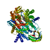
Mass: 393.418 Da / Num. of mol.: 1 / Source method: obtained synthetically / Formula: C20H20FN7O / Feature type: SUBJECT OF INVESTIGATION
Has ligand of interestY

-
Experimental details

-
Experiment

ExperimentMethod: X-RAY DIFFRACTION / Number of used crystals: 1

-
Sample preparation

CrystalDensity Matthews: 2.34 Å3/Da / Density % sol: 47.51 % / Mosaicity: 0 °
Crystal growTemperature: 293 K / Method: vapor diffusion / pH: 8
Details: 20 % PEG3350, 0.175 M ammonium sulfate, 0.001 M TCEP, 0.1 M Hepes pH 8.0

-
Data collection

DiffractionMean temperature: 100 K / Serial crystal experiment: N
Diffraction sourceSource: SYNCHROTRON / Site: Diamond / Beamline: I03 / Wavelength: 0.97625 Å
DetectorType: DECTRIS PILATUS 6M / Detector: PIXEL / Date: Apr 27, 2019
RadiationProtocol: SINGLE WAVELENGTH / Monochromatic (M) / Laue (L): M / Scattering type: x-ray
Radiation wavelengthWavelength: 0.97625 Å / Relative weight: 1
ReflectionResolution: 2.738→62.21 Å / Num. obs: 26986 / % possible obs: 99.2 % / Redundancy: 3.3 % / Biso Wilson estimate: 129.48 Å2 / CC1/2: 0.999 / Rmerge(I) obs: 0.037 / Rpim(I) all: 0.024 / Rrim(I) all: 0.045 / Net I/σ(I): 11.2
Reflection shell

Diffraction-ID: 1

Resolution (Å)Redundancy (%)Rmerge(I) obsNum. measured allNum. unique obsCC1/2Rpim(I) allRrim(I) allNet I/σ(I) obs% possible all
2.74-2.813.40.796675419800.6030.5020.944199.3
12.25-62.1630.0218082720.9960.0140.02530.381.1

-
Phasing

PhasingMethod: molecular replacement

-
Processing

Software
NameVersionClassification
Aimless0.7.4data scaling
AMoREphasing
BUSTER2.11.8 (24-FEB-2021)refinement
PDB_EXTRACT3.27data extraction
DIALSdata reduction
RefinementMethod to determine structure: MOLECULAR REPLACEMENT
Starting model: internal model

Resolution: 2.738→62.21 Å / Cor.coef. Fo:Fc: 0.955 / Cor.coef. Fo:Fc free: 0.935 / Cross valid method: THROUGHOUT / σ(F): 0 / SU R Blow DPI: 1.51 / SU Rfree Blow DPI: 0.364
RfactorNum. reflection% reflectionSelection details
Rfree0.2757 1317 4.88 %RANDOM
Rwork0.2174 ---
obs0.2203 26976 98.9 %-
Displacement parametersBiso max: 220.39 Å2 / Biso mean: 141.29 Å2 / Biso min: 80.6 Å2
Baniso -1Baniso -2Baniso -3
1-7.3619 Å20 Å20.5769 Å2
2---10.489 Å20 Å2
3---3.1271 Å2
Refine analyzeLuzzati coordinate error obs: 0.39 Å
Refinement stepCycle: final / Resolution: 2.738→62.21 Å
ProteinNucleic acidLigandSolventTotal
Num. atoms6749 0 29 0 6778
Biso mean--109.33 --
Num. residues----834
Refine LS restraints
Refine-IDTypeNumberRestraint functionWeightDev ideal
X-RAY DIFFRACTIONt_dihedral_angle_d2452SINUSOIDAL2
X-RAY DIFFRACTIONt_trig_c_planes
X-RAY DIFFRACTIONt_gen_planes1153HARMONIC5
X-RAY DIFFRACTIONt_it6927HARMONIC10
X-RAY DIFFRACTIONt_nbd
X-RAY DIFFRACTIONt_improper_torsion
X-RAY DIFFRACTIONt_pseud_angle
X-RAY DIFFRACTIONt_chiral_improper_torsion900SEMIHARMONIC5
X-RAY DIFFRACTIONt_sum_occupancies
X-RAY DIFFRACTIONt_utility_distance
X-RAY DIFFRACTIONt_utility_angle
X-RAY DIFFRACTIONt_utility_torsion
X-RAY DIFFRACTIONt_ideal_dist_contact5130SEMIHARMONIC4
X-RAY DIFFRACTIONt_bond_d6927HARMONIC20.009
X-RAY DIFFRACTIONt_angle_deg9377HARMONIC21
X-RAY DIFFRACTIONt_omega_torsion2.72
X-RAY DIFFRACTIONt_other_torsion20.65
LS refinement shellResolution: 2.74→2.76 Å / Rfactor Rfree error: 0 / Total num. of bins used: 51
RfactorNum. reflection% reflection
Rfree0.3066 30 5.56 %
Rwork0.3204 510 -
all0.3196 540 -
obs--90.74 %
Refinement TLS params.Method: refined / Origin x: 32.3272 Å / Origin y: -0.014 Å / Origin z: 26.2773 Å
111213212223313233
T-0.1958 Å20.0205 Å20.0679 Å2--0.5091 Å2-0.0399 Å2---0.2561 Å2
L7.2362 °21.2366 °21.2433 °2-1.5413 °20.065 °2--3.8571 °2
S0.0473 Å °-0.1307 Å °-0.0404 Å °-0.0789 Å °0.0174 Å °-0.3617 Å °-0.5213 Å °0.1334 Å °-0.0647 Å °
Refinement TLS groupSelection details: { A|* }

+
About Yorodumi

-
News

-
Feb 9, 2022. New format data for meta-information of EMDB entries

New format data for meta-information of EMDB entries

  • Version 3 of the EMDB header file is now the official format.
  • The previous official version 1.9 will be removed from the archive.

Related info.:EMDB header

External links:wwPDB to switch to version 3 of the EMDB data model

-
Aug 12, 2020. Covid-19 info

Covid-19 info

URL: https://pdbj.org/emnavi/covid19.php

New page: Covid-19 featured information page in EM Navigator.

Related info.:Covid-19 info / Mar 5, 2020. Novel coronavirus structure data

+
Mar 5, 2020. Novel coronavirus structure data

Novel coronavirus structure data

Related info.:Yorodumi Speices / Aug 12, 2020. Covid-19 info

External links:COVID-19 featured content - PDBj / Molecule of the Month (242):Coronavirus Proteases

+
Jan 31, 2019. EMDB accession codes are about to change! (news from PDBe EMDB page)

EMDB accession codes are about to change! (news from PDBe EMDB page)

  • The allocation of 4 digits for EMDB accession codes will soon come to an end. Whilst these codes will remain in use, new EMDB accession codes will include an additional digit and will expand incrementally as the available range of codes is exhausted. The current 4-digit format prefixed with “EMD-” (i.e. EMD-XXXX) will advance to a 5-digit format (i.e. EMD-XXXXX), and so on. It is currently estimated that the 4-digit codes will be depleted around Spring 2019, at which point the 5-digit format will come into force.
  • The EM Navigator/Yorodumi systems omit the EMD- prefix.

Related info.:Q: What is EMD? / ID/Accession-code notation in Yorodumi/EM Navigator

External links:EMDB Accession Codes are Changing Soon! / Contact to PDBj

+
Jul 12, 2017. Major update of PDB

Major update of PDB

  • wwPDB released updated PDB data conforming to the new PDBx/mmCIF dictionary.
  • This is a major update changing the version number from 4 to 5, and with Remediation, in which all the entries are updated.
  • In this update, many items about electron microscopy experimental information are reorganized (e.g. em_software).
  • Now, EM Navigator and Yorodumi are based on the updated data.

External links:wwPDB Remediation / Enriched Model Files Conforming to OneDep Data Standards Now Available in the PDB FTP Archive

-
Yorodumi

Thousand views of thousand structures

  • Yorodumi is a browser for structure data from EMDB, PDB, SASBDB, etc.
  • This page is also the successor to EM Navigator detail page, and also detail information page/front-end page for Omokage search.
  • The word "yorodu" (or yorozu) is an old Japanese word meaning "ten thousand". "mi" (miru) is to see.

Related info.:EMDB / PDB / SASBDB / Comparison of 3 databanks / Yorodumi Search / Aug 31, 2016. New EM Navigator & Yorodumi / Yorodumi Papers / Jmol/JSmol / Function and homology information / Changes in new EM Navigator and Yorodumi

Read more