 Movie
Movie Controller

[English] 日本語
 Yorodumi
- PDB-7z0e: Crystal structure of the M state of bacteriorhodopsin at 1.22 Ang... -
+ Open data

- Basic information
| Entry | Database: PDB / ID: 7z0e | ||||||||||||||||||
|---|---|---|---|---|---|---|---|---|---|---|---|---|---|---|---|---|---|---|---|
| Title | Crystal structure of the M state of bacteriorhodopsin at 1.22 Angstrom resolution | ||||||||||||||||||
 Components Components | Bacteriorhodopsin | ||||||||||||||||||
 Keywords Keywords | MEMBRANE PROTEIN / rhodopsin / retinal / microbial rhodopsin / ion transport / proton pump / photocycle / ultrahigh resolution | ||||||||||||||||||
| Function / homology |  Function and homology informationphotoreceptor activity / phototransduction / proton transmembrane transport / monoatomic ion channel activity / plasma membrane Function and homology informationphotoreceptor activity / phototransduction / proton transmembrane transport / monoatomic ion channel activity / plasma membraneSimilarity search - Function | ||||||||||||||||||
| Biological species |  Halobacterium salinarum (Halophile) Halobacterium salinarum (Halophile) | ||||||||||||||||||
| Method | X-RAY DIFFRACTION / SYNCHROTRON / MOLECULAR REPLACEMENT / Resolution: 1.22 Å | ||||||||||||||||||
 Authors Authors | Borshchevskiy, V. / Kovalev, K. / Round, E. / Efremov, R. / Bourenkov, G. / Gordeliy, V. | ||||||||||||||||||
| Funding support |  France, France,  Russian Federation, 5items Russian Federation, 5items
| ||||||||||||||||||
 Citation Citation | Journal: Nat.Struct.Mol.Biol. / Year: 2022 Title: True-atomic-resolution insights into the structure and functional role of linear chains and low-barrier hydrogen bonds in proteins. Authors: Borshchevskiy, V. / Kovalev, K. / Round, E. / Efremov, R. / Astashkin, R. / Bourenkov, G. / Bratanov, D. / Balandin, T. / Chizhov, I. / Baeken, C. / Gushchin, I. / Kuzmin, A. / Alekseev, A. ...Authors: Borshchevskiy, V. / Kovalev, K. / Round, E. / Efremov, R. / Astashkin, R. / Bourenkov, G. / Bratanov, D. / Balandin, T. / Chizhov, I. / Baeken, C. / Gushchin, I. / Kuzmin, A. / Alekseev, A. / Rogachev, A. / Willbold, D. / Engelhard, M. / Bamberg, E. / Buldt, G. / Gordeliy, V. | ||||||||||||||||||
| History |
|
- Structure visualization
| Structure viewer | Molecule: MolmilJmol/JSmol |
|---|
- Downloads & links
-Download
| PDBx/mmCIF format | 7z0e.cif.gz | 220.8 KB | Display | PDBx/mmCIF format |
|---|---|---|---|---|
| PDB format | pdb7z0e.ent.gz | 187.4 KB | Display | PDB format |
| PDBx/mmJSON format | 7z0e.json.gz | Tree view | PDBx/mmJSON format | |
| Others |  Other downloads Other downloads |
-Validation report
| Arichive directory | https://data.pdbj.org/pub/pdb/validation_reports/z0/7z0eftp://data.pdbj.org/pub/pdb/validation_reports/z0/7z0e | HTTPS FTP |
|---|
-Related structure data
| Related structure data |  7z09C 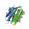 7z0aC 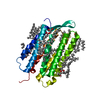 7z0cC 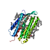 7z0dC 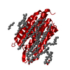 1c3wS S: Starting model for refinement C: citing same article ( |
|---|---|
| Similar structure data |
-Links
 PDBj
PDBj


- Assembly
| Deposited unit | 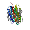
| ||||||||
|---|---|---|---|---|---|---|---|---|---|
| 1 |
| ||||||||
| Unit cell |
|
-Components
-Protein , 1 types, 1 molecules P
| #1: Protein | / BR / Bacterioopsin / BO Mass: 27081.840 Da / Num. of mol.: 1 / Source method: isolated from a natural source / Source: (natural) Halobacterium salinarum (Halophile) / Strain: ATCC 700922 / JCM 11081 / NRC-1 / References: UniProt: P02945 |
|---|
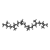
-Non-polymers , 5 types, 96 molecules 

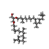

| #2: Chemical | ChemComp-OLC / ( Mass: 356.540 Da / Num. of mol.: 1 / Source method: obtained synthetically / Formula: C21H40O4 Mass: 356.540 Da / Num. of mol.: 1 / Source method: obtained synthetically / Formula: C21H40O4 | ||||||
|---|---|---|---|---|---|---|---|
| #3: Chemical | ChemComp-LFA / Icosane Mass: 282.547 Da / Num. of mol.: 19 / Source method: obtained synthetically / Formula: C20H42 Mass: 282.547 Da / Num. of mol.: 19 / Source method: obtained synthetically / Formula: C20H42#4: Chemical | ChemComp-L2P / |  Mass: 653.157 Da / Num. of mol.: 1 / Source method: obtained synthetically / Formula: C43H88O3 Mass: 653.157 Da / Num. of mol.: 1 / Source method: obtained synthetically / Formula: C43H88O3#5: Chemical | ChemComp-SQL / ( | Squalene Mass: 410.718 Da / Num. of mol.: 1 / Source method: obtained synthetically / Formula: C30H50 Mass: 410.718 Da / Num. of mol.: 1 / Source method: obtained synthetically / Formula: C30H50#6: Water | ChemComp-HOH / | WaterMass: 18.015 Da / Num. of mol.: 74 / Source method: isolated from a natural source / Formula: H2O |
-Details
| Has ligand of interest | Y |
|---|
-Experimental details
-Experiment
| Experiment | Method: X-RAY DIFFRACTION / Number of used crystals: 1 |
|---|
- Sample preparation
| Crystal | Density Matthews: 2.19 Å3/Da / Density % sol: 43.85 % |
|---|---|
| Crystal grow | Temperature: 293 K / Method: lipidic cubic phase / Details: Na2HPO4 (5%) and KH2PO4 (95%) |
-Data collection
| Diffraction | Mean temperature: 100 K / Serial crystal experiment: N | ||||||||||||||||||||||||||||||||||||||||||||||||||||||||||||||||||||||||||||||||||||||||||||||||||||||||||||||||||||||||||||||||||||||||||||||||||||||||||||||||||||||||||||||||||||||||||||||||||||||||||||||||||
|---|---|---|---|---|---|---|---|---|---|---|---|---|---|---|---|---|---|---|---|---|---|---|---|---|---|---|---|---|---|---|---|---|---|---|---|---|---|---|---|---|---|---|---|---|---|---|---|---|---|---|---|---|---|---|---|---|---|---|---|---|---|---|---|---|---|---|---|---|---|---|---|---|---|---|---|---|---|---|---|---|---|---|---|---|---|---|---|---|---|---|---|---|---|---|---|---|---|---|---|---|---|---|---|---|---|---|---|---|---|---|---|---|---|---|---|---|---|---|---|---|---|---|---|---|---|---|---|---|---|---|---|---|---|---|---|---|---|---|---|---|---|---|---|---|---|---|---|---|---|---|---|---|---|---|---|---|---|---|---|---|---|---|---|---|---|---|---|---|---|---|---|---|---|---|---|---|---|---|---|---|---|---|---|---|---|---|---|---|---|---|---|---|---|---|---|---|---|---|---|---|---|---|---|---|---|---|---|---|---|---|---|
| Diffraction source | Source: SYNCHROTRON / Site: ESRF / Beamline: ID14-1 / Wavelength: 0.934 Å | ||||||||||||||||||||||||||||||||||||||||||||||||||||||||||||||||||||||||||||||||||||||||||||||||||||||||||||||||||||||||||||||||||||||||||||||||||||||||||||||||||||||||||||||||||||||||||||||||||||||||||||||||||
| Detector | Type: ADSC QUANTUM 210 / Detector: CCD / Date: Jul 10, 2006 | ||||||||||||||||||||||||||||||||||||||||||||||||||||||||||||||||||||||||||||||||||||||||||||||||||||||||||||||||||||||||||||||||||||||||||||||||||||||||||||||||||||||||||||||||||||||||||||||||||||||||||||||||||
| Radiation | Protocol: SINGLE WAVELENGTH / Monochromatic (M) / Laue (L): M / Scattering type: x-ray | ||||||||||||||||||||||||||||||||||||||||||||||||||||||||||||||||||||||||||||||||||||||||||||||||||||||||||||||||||||||||||||||||||||||||||||||||||||||||||||||||||||||||||||||||||||||||||||||||||||||||||||||||||
| Radiation wavelength | Wavelength: 0.934 Å / Relative weight: 1 | ||||||||||||||||||||||||||||||||||||||||||||||||||||||||||||||||||||||||||||||||||||||||||||||||||||||||||||||||||||||||||||||||||||||||||||||||||||||||||||||||||||||||||||||||||||||||||||||||||||||||||||||||||
| Reflection twin | Operator: k,h,-l / Fraction: 0.32 | ||||||||||||||||||||||||||||||||||||||||||||||||||||||||||||||||||||||||||||||||||||||||||||||||||||||||||||||||||||||||||||||||||||||||||||||||||||||||||||||||||||||||||||||||||||||||||||||||||||||||||||||||||
| Reflection | Resolution: 1.22→30.445 Å / Num. obs: 66766 / % possible obs: 98.1 % / Redundancy: 6.407 % / CC1/2: 0.999 / Rmerge(I) obs: 0.066 / Rrim(I) all: 0.071 / Χ2: 0.963 / Net I/σ(I): 13.75 / Num. measured all: 427782 / Scaling rejects: 118 | ||||||||||||||||||||||||||||||||||||||||||||||||||||||||||||||||||||||||||||||||||||||||||||||||||||||||||||||||||||||||||||||||||||||||||||||||||||||||||||||||||||||||||||||||||||||||||||||||||||||||||||||||||
| Reflection shell | Diffraction-ID: 1
|
- Processing
| Software |
| ||||||||||||||||||
|---|---|---|---|---|---|---|---|---|---|---|---|---|---|---|---|---|---|---|---|
| Refinement | Method to determine structure: MOLECULAR REPLACEMENT Starting model: 1C3W Resolution: 1.22→30.445 Å / Cross valid method: THROUGHOUT
| ||||||||||||||||||
| Displacement parameters | Biso max: 120.43 Å2 / Biso mean: 28.9432 Å2 / Biso min: 10.21 Å2 | ||||||||||||||||||
| Refinement step | Cycle: LAST / Resolution: 1.22→30.445 Å
| ||||||||||||||||||
| LS refinement shell | Resolution: 1.2204→1.2487 Å
|
