 Movie
Movie Controller

[English] 日本語
 Yorodumi
- PDB-7yva: Crystal structure of Candida albicans Fructose-1,6-bisphosphate a... -
+ Open data

- Basic information
| Entry | Database: PDB / ID: 7yva | ||||||
|---|---|---|---|---|---|---|---|
| Title | Crystal structure of Candida albicans Fructose-1,6-bisphosphate aldolase complexed with lipoic acid | ||||||
 Components Components | Candida albicans Fructose-1,6-bisphosphate aldolase | ||||||
 Keywords Keywords |  CYTOSOLIC PROTEIN/INHIBITOR / non-covalent inhibitor / CYTOSOLIC PROTEIN-INHIBITOR COMPLEX CYTOSOLIC PROTEIN/INHIBITOR / non-covalent inhibitor / CYTOSOLIC PROTEIN-INHIBITOR COMPLEX | ||||||
| Function / homology |  Function and homology information: / hyphal cell wall / fungal-type cell wall / fructose-bisphosphate aldolase / fructose-bisphosphate aldolase activity / biological process involved in interaction with host / gluconeogenesis / glycolytic process / cell surface / zinc ion binding ...: / hyphal cell wall / fungal-type cell wall / fructose-bisphosphate aldolase / fructose-bisphosphate aldolase activity / biological process involved in interaction with host / gluconeogenesis / glycolytic process / cell surface / zinc ion binding / plasma membrane / cytosol Function and homology information: / hyphal cell wall / fungal-type cell wall / fructose-bisphosphate aldolase / fructose-bisphosphate aldolase activity / biological process involved in interaction with host / gluconeogenesis / glycolytic process / cell surface / zinc ion binding ...: / hyphal cell wall / fungal-type cell wall / fructose-bisphosphate aldolase / fructose-bisphosphate aldolase activity / biological process involved in interaction with host / gluconeogenesis / glycolytic process / cell surface / zinc ion binding / plasma membrane / cytosolSimilarity search - Function | ||||||
| Biological species |  Candida albicans (yeast) Candida albicans (yeast) | ||||||
| Method | X-RAY DIFFRACTION / SYNCHROTRON / MOLECULAR REPLACEMENT / Resolution: 2.93 Å | ||||||
 Authors Authors | Cao, H. / Huang, Y. / Ren, Y. / Wan, J. | ||||||
| Funding support |  China, 1items China, 1items
| ||||||
 Citation Citation | Journal: To Be Published Title: Crystal structure of Candida albicans Fructose-1,6-bisphosphate aldolase complexed with lipoic acid Authors: Cao, H. / Huang, Y. / Ren, Y. / Wan, J. | ||||||
| History |
|
- Structure visualization
| Structure viewer | Molecule: MolmilJmol/JSmol |
|---|
- Downloads & links
-Download
| PDBx/mmCIF format | 7yva.cif.gz | 276.5 KB | Display | PDBx/mmCIF format |
|---|---|---|---|---|
| PDB format | pdb7yva.ent.gz | 223.6 KB | Display | PDB format |
| PDBx/mmJSON format | 7yva.json.gz | Tree view | PDBx/mmJSON format | |
| Others |  Other downloads Other downloads |
-Validation report
| Arichive directory | https://data.pdbj.org/pub/pdb/validation_reports/yv/7yvaftp://data.pdbj.org/pub/pdb/validation_reports/yv/7yva | HTTPS FTP |
|---|
-Related structure data
| Related structure data | 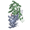 6lnkS S: Starting model for refinement |
|---|---|
| Similar structure data |
-Links
 PDBj
PDBj
- Assembly
| Deposited unit | 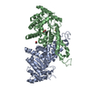
| ||||||||
|---|---|---|---|---|---|---|---|---|---|
| 1 |
| ||||||||
| Unit cell |
|
-Components
| #1: Protein | Mass: 39263.273 Da / Num. of mol.: 2 Source method: isolated from a genetically manipulated source Source: (gene. exp.) Candida albicans (yeast)Production host:  Escherichia coli 'BL21-Gold(DE3)pLysS AG' (bacteria) Escherichia coli 'BL21-Gold(DE3)pLysS AG' (bacteria)References: UniProt: Q9URB4 #2: Chemical |   Mass: 65.409 Da / Num. of mol.: 2 / Source method: obtained synthetically / Formula: Zn Mass: 65.409 Da / Num. of mol.: 2 / Source method: obtained synthetically / Formula: Zn#3: Chemical | ChemComp-LPA / | Lipoic acid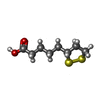  Mass: 206.326 Da / Num. of mol.: 1 / Source method: obtained synthetically / Formula: C8H14O2S2 / Feature type: SUBJECT OF INVESTIGATION Mass: 206.326 Da / Num. of mol.: 1 / Source method: obtained synthetically / Formula: C8H14O2S2 / Feature type: SUBJECT OF INVESTIGATION#4: Chemical | Ethylene glycol  Mass: 62.068 Da / Num. of mol.: 2 / Source method: obtained synthetically / Formula: C2H6O2 Mass: 62.068 Da / Num. of mol.: 2 / Source method: obtained synthetically / Formula: C2H6O2#5: Water | ChemComp-HOH / | Water Mass: 18.015 Da / Num. of mol.: 103 / Source method: isolated from a natural source / Formula: H2O Mass: 18.015 Da / Num. of mol.: 103 / Source method: isolated from a natural source / Formula: H2OHas ligand of interest | Y | |
|---|
-Experimental details
-Experiment
| Experiment | Method: X-RAY DIFFRACTION / Number of used crystals: 1 |
|---|
- Sample preparation
| Crystal | Density Matthews: 2.33 Å3/Da / Density % sol: 47.3 % |
|---|---|
| Crystal grow | Temperature: 291 K / Method: vapor diffusion, hanging drop / Details: 22.5% (w/v) PEG 3350 100 mM Hepes pH =7.6 |
-Data collection
| Diffraction | Mean temperature: 100 K / Serial crystal experiment: N |
|---|---|
| Diffraction source | Source: SYNCHROTRON / Site: SSRF / Beamline: BL19U1 / Wavelength: 0.98 Å |
| Detector | Type: DECTRIS PILATUS 6M / Detector: PIXEL / Date: Oct 30, 2019 |
| Radiation | Protocol: SINGLE WAVELENGTH / Monochromatic (M) / Laue (L): M / Scattering type: x-ray |
| Radiation wavelength | Wavelength: 0.98 Å / Relative weight: 1 |
| Reflection | Resolution: 2.93→45.37 Å / Num. obs: 15544 / % possible obs: 98.47 % / Redundancy: 3.3 % / Biso Wilson estimate: 79.64 Å2 / CC1/2: 0.998 / Net I/σ(I): 9.38 |
| Reflection shell | Resolution: 2.93→3.04 Å / Num. unique obs: 1493 / CC1/2: 0.752 |
- Processing
| Software |
| ||||||||||||||||||||||||||||||||||||||||||||||||||||||||||||||||||||||||||||||||||||
|---|---|---|---|---|---|---|---|---|---|---|---|---|---|---|---|---|---|---|---|---|---|---|---|---|---|---|---|---|---|---|---|---|---|---|---|---|---|---|---|---|---|---|---|---|---|---|---|---|---|---|---|---|---|---|---|---|---|---|---|---|---|---|---|---|---|---|---|---|---|---|---|---|---|---|---|---|---|---|---|---|---|---|---|---|---|
| Refinement | Method to determine structure: MOLECULAR REPLACEMENT Starting model: 6LNK Resolution: 2.93→45.37 Å / SU ML: 0.43 / Cross valid method: THROUGHOUT / σ(F): 1.38 / Phase error: 27.7 / Stereochemistry target values: ML
| ||||||||||||||||||||||||||||||||||||||||||||||||||||||||||||||||||||||||||||||||||||
| Solvent computation | Shrinkage radii: 0.9 Å / VDW probe radii: 1.1 Å / Solvent model: FLAT BULK SOLVENT MODEL | ||||||||||||||||||||||||||||||||||||||||||||||||||||||||||||||||||||||||||||||||||||
| Displacement parameters | Biso max: 152.36 Å2 / Biso mean: 77.9798 Å2 / Biso min: 47.79 Å2 | ||||||||||||||||||||||||||||||||||||||||||||||||||||||||||||||||||||||||||||||||||||
| Refinement step | Cycle: final / Resolution: 2.93→45.37 Å
| ||||||||||||||||||||||||||||||||||||||||||||||||||||||||||||||||||||||||||||||||||||
| LS refinement shell | Refine-ID: X-RAY DIFFRACTION / Rfactor Rfree error: 0 / Total num. of bins used: 11
| ||||||||||||||||||||||||||||||||||||||||||||||||||||||||||||||||||||||||||||||||||||
| Refinement TLS params. | Method: refined / Origin x: -23.2191 Å / Origin y: 1.4468 Å / Origin z: 17.3697 Å
| ||||||||||||||||||||||||||||||||||||||||||||||||||||||||||||||||||||||||||||||||||||
| Refinement TLS group |
|
