 Movie
Movie Controller


+ Open data

- Basic information
| Entry | Database: PDB / ID: 7yr9 | ||||||
|---|---|---|---|---|---|---|---|
| Title | Crystal structure of the immature form of TtPetA | ||||||
 Components Components | Ubiquinol-cytochrome c reductase iron-sulfur subunit | ||||||
 Keywords Keywords |  ELECTRON TRANSPORT / Rieske / iron-sulfur cluster / metal binding ELECTRON TRANSPORT / Rieske / iron-sulfur cluster / metal binding | ||||||
| Function / homology |  Function and homology information Function and homology informationquinol-cytochrome-c reductase / ubiquinol-cytochrome-c reductase activity / 2 iron, 2 sulfur cluster binding / intracellular membrane-bounded organelle / metal ion binding / plasma membrane / cytoplasmSimilarity search - Function | ||||||
| Biological species |  Thermochromatium tepidum (bacteria) Thermochromatium tepidum (bacteria) | ||||||
| Method | X-RAY DIFFRACTION / SYNCHROTRON / SAD / Resolution: 1.7 Å | ||||||
 Authors Authors | Tsutsumi, E. / Niwa, S. / Takeda, K. | ||||||
| Funding support |  Japan, 1items Japan, 1items
| ||||||
 Citation Citation | Journal: Commun Chem / Year: 2023 Title: Structure of a putative immature form of a Rieske-type iron-sulfur protein in complex with zinc chloride. Authors: Tsutsumi, E. / Niwa, S. / Takeda, R. / Sakamoto, N. / Okatsu, K. / Fukai, S. / Ago, H. / Nagao, S. / Sekiguchi, H. / Takeda, K. | ||||||
| History |
|
- Structure visualization
| Structure viewer | Molecule: MolmilJmol/JSmol |
|---|
- Downloads & links
-Download
| PDBx/mmCIF format | 7yr9.cif.gz | 76.5 KB | Display | PDBx/mmCIF format |
|---|---|---|---|---|
| PDB format | pdb7yr9.ent.gz | 53.9 KB | Display | PDB format |
| PDBx/mmJSON format | 7yr9.json.gz | Tree view | PDBx/mmJSON format | |
| Others |  Other downloads Other downloads |
-Validation report
| Arichive directory | https://data.pdbj.org/pub/pdb/validation_reports/yr/7yr9ftp://data.pdbj.org/pub/pdb/validation_reports/yr/7yr9 | HTTPS FTP |
|---|
-Related structure data
| Related structure data |  7yraC C: citing same article ( |
|---|---|
| Similar structure data | |
| Other databases |
|
-Links
 PDBj
PDBj


- Assembly
| Deposited unit | 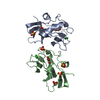
| ||||||||
|---|---|---|---|---|---|---|---|---|---|
| 1 | 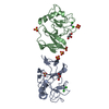
| ||||||||
| Unit cell |
|
-Components
-Protein , 1 types, 2 molecules AB
| #1: Protein | Mass: 16449.885 Da / Num. of mol.: 2 Source method: isolated from a genetically manipulated source Source: (gene. exp.) Thermochromatium tepidum (bacteria) / Gene: petA / Production host: Escherichia coli (E. coli) / References: UniProt: D1MZ11, quinol-cytochrome-c reductase |
|---|
-Non-polymers , 5 types, 159 molecules 


| #2: Chemical |  Mass: 65.409 Da / Num. of mol.: 2 / Source method: obtained synthetically / Formula: Zn / Feature type: SUBJECT OF INVESTIGATION Mass: 65.409 Da / Num. of mol.: 2 / Source method: obtained synthetically / Formula: Zn / Feature type: SUBJECT OF INVESTIGATION#3: Chemical | ChemComp-CL / Chloride Mass: 35.453 Da / Num. of mol.: 4 / Source method: obtained synthetically / Formula: Cl / Feature type: SUBJECT OF INVESTIGATION Mass: 35.453 Da / Num. of mol.: 4 / Source method: obtained synthetically / Formula: Cl / Feature type: SUBJECT OF INVESTIGATION#4: Chemical | ChemComp-SO4 / Sulfate Mass: 96.063 Da / Num. of mol.: 6 / Source method: obtained synthetically / Formula: SO4 Mass: 96.063 Da / Num. of mol.: 6 / Source method: obtained synthetically / Formula: SO4#5: Chemical | Glycerol Mass: 92.094 Da / Num. of mol.: 2 / Source method: obtained synthetically / Formula: C3H8O3 Mass: 92.094 Da / Num. of mol.: 2 / Source method: obtained synthetically / Formula: C3H8O3#6: Water | ChemComp-HOH / | WaterMass: 18.015 Da / Num. of mol.: 145 / Source method: isolated from a natural source / Formula: H2O |
|---|
-Details
| Has ligand of interest | Y |
|---|
-Experimental details
-Experiment
| Experiment | Method: X-RAY DIFFRACTION / Number of used crystals: 1 |
|---|
- Sample preparation
| Crystal | Density Matthews: 2.51 Å3/Da / Density % sol: 50.93 % |
|---|---|
| Crystal grow | Temperature: 293 K / Method: vapor diffusion, sitting drop / Details: 1.6 M AmS, 20% glycerol, 0.1 M Na-Ac pH 4.6 |
-Data collection
| Diffraction | Mean temperature: 15 K / Ambient temp details: He cryostream / Serial crystal experiment: N | ||||||||||||||||||||||||||||||||||||||||||||||||||||||||||||||||||||||||||||||||||||||||||||||||||||
|---|---|---|---|---|---|---|---|---|---|---|---|---|---|---|---|---|---|---|---|---|---|---|---|---|---|---|---|---|---|---|---|---|---|---|---|---|---|---|---|---|---|---|---|---|---|---|---|---|---|---|---|---|---|---|---|---|---|---|---|---|---|---|---|---|---|---|---|---|---|---|---|---|---|---|---|---|---|---|---|---|---|---|---|---|---|---|---|---|---|---|---|---|---|---|---|---|---|---|---|---|---|
| Diffraction source | Source: SYNCHROTRON / Site: SPring-8 / Beamline: BL41XU / Wavelength: 1.2825 Å | ||||||||||||||||||||||||||||||||||||||||||||||||||||||||||||||||||||||||||||||||||||||||||||||||||||
| Detector | Type: DECTRIS EIGER X 16M / Detector: PIXEL / Date: Jul 25, 2020 | ||||||||||||||||||||||||||||||||||||||||||||||||||||||||||||||||||||||||||||||||||||||||||||||||||||
| Radiation | Protocol: SINGLE WAVELENGTH / Monochromatic (M) / Laue (L): M / Scattering type: x-ray | ||||||||||||||||||||||||||||||||||||||||||||||||||||||||||||||||||||||||||||||||||||||||||||||||||||
| Radiation wavelength | Wavelength: 1.2825 Å / Relative weight: 1 | ||||||||||||||||||||||||||||||||||||||||||||||||||||||||||||||||||||||||||||||||||||||||||||||||||||
| Reflection twin | Operator: -h,-k,l / Fraction: 0.256 | ||||||||||||||||||||||||||||||||||||||||||||||||||||||||||||||||||||||||||||||||||||||||||||||||||||
| Reflection | Resolution: 1.7→50 Å / Num. obs: 34104 / % possible obs: 95.7 % / Redundancy: 7.09 % / Biso Wilson estimate: 21.741 Å2 / CC1/2: 0.999 / Rmerge(I) obs: 0.078 / Rrim(I) all: 0.084 / Χ2: 0.9 / Net I/σ(I): 15.94 | ||||||||||||||||||||||||||||||||||||||||||||||||||||||||||||||||||||||||||||||||||||||||||||||||||||
| Reflection shell | Diffraction-ID: 1
|
-Phasing
| Phasing | Method: SAD | ||||||||||||||||||||||||||||||||||||||||||||||||||||||||||||||||||||||||||||||||||||||||||||||||||||||||||||||||||||
|---|---|---|---|---|---|---|---|---|---|---|---|---|---|---|---|---|---|---|---|---|---|---|---|---|---|---|---|---|---|---|---|---|---|---|---|---|---|---|---|---|---|---|---|---|---|---|---|---|---|---|---|---|---|---|---|---|---|---|---|---|---|---|---|---|---|---|---|---|---|---|---|---|---|---|---|---|---|---|---|---|---|---|---|---|---|---|---|---|---|---|---|---|---|---|---|---|---|---|---|---|---|---|---|---|---|---|---|---|---|---|---|---|---|---|---|---|---|
| Phasing dm | Method: Solvent flattening and Histogram matching / Reflection: 33923 | ||||||||||||||||||||||||||||||||||||||||||||||||||||||||||||||||||||||||||||||||||||||||||||||||||||||||||||||||||||
| Phasing dm shell |
|
- Processing
| Software |
| ||||||||||||||||||||||||
|---|---|---|---|---|---|---|---|---|---|---|---|---|---|---|---|---|---|---|---|---|---|---|---|---|---|
| Refinement | Method to determine structure: SAD / Resolution: 1.7→50 Å / Cross valid method: THROUGHOUT / σ(F): 2885
| ||||||||||||||||||||||||
| Solvent computation | Bsol: 57.4944 Å2 | ||||||||||||||||||||||||
| Displacement parameters | Biso max: 75 Å2 / Biso mean: 15.9042 Å2 / Biso min: 4.65 Å2
| ||||||||||||||||||||||||
| Refinement step | Cycle: final / Resolution: 1.7→50 Å
| ||||||||||||||||||||||||
| Refine LS restraints |
| ||||||||||||||||||||||||
| LS refinement shell | Resolution: 1.7→1.78 Å
| ||||||||||||||||||||||||
| Xplor file |
|
