 Movie
Movie Controller

[English] 日本語
 Yorodumi
- PDB-7yom: Crystal structure of tetra mutant (D67E,A68P,L98I,A301S) of O-ace... -
+ Open data

- Basic information
| Entry | Database: PDB / ID: 7yom | ||||||
|---|---|---|---|---|---|---|---|
| Title | Crystal structure of tetra mutant (D67E,A68P,L98I,A301S) of O-acetylserine sulfhydrylase from Salmonella typhimurium in complex with high-affinity inhibitory peptide from serine acetyltransferase of Salmonella typhimurium at 2.8 A | ||||||
 Components Components |
| ||||||
 Keywords Keywords | TRANSFERASE/TRANSFERASE INHIBITOR / tetra mutant (D67E / A68P / L98I / A301S) / O-acetylserine sulfhydrylase /  Salmonella typhimurium / high-affinity inhibitory peptide from serine acetyltransferase / TRANSFERASE-TRANSFERASE INHIBITOR complex Salmonella typhimurium / high-affinity inhibitory peptide from serine acetyltransferase / TRANSFERASE-TRANSFERASE INHIBITOR complex | ||||||
| Function / homology |  Function and homology informationcysteine synthase / cystathionine beta-synthase activity / cysteine synthase activity / L-cysteine desulfhydrase activity / cysteine biosynthetic process from serine / cytoplasm Function and homology informationcysteine synthase / cystathionine beta-synthase activity / cysteine synthase activity / L-cysteine desulfhydrase activity / cysteine biosynthetic process from serine / cytoplasmSimilarity search - Function | ||||||
| Biological species |  Haemophilus influenzae Rd KW20 (bacteria)Salmonella typhimurium (bacteria) Haemophilus influenzae Rd KW20 (bacteria)Salmonella typhimurium (bacteria) | ||||||
| Method | X-RAY DIFFRACTION / MOLECULAR REPLACEMENT / Resolution: 2.8 Å | ||||||
 Authors Authors | Saini, N. / Kumar, N. / Rahisuddin, R. / Singh, A.K. | ||||||
| Funding support |  India, 1items India, 1items
| ||||||
 Citation Citation | Journal: To Be Published Title: Crystal structure of I88L single mutant of O-acetylserine sulfhydrylase from Haemophilus influenzae in complex with high-affinity inhibitory peptide from serine acetyltransferase of Salmonella ...Title: Crystal structure of I88L single mutant of O-acetylserine sulfhydrylase from Haemophilus influenzae in complex with high-affinity inhibitory peptide from serine acetyltransferase of Salmonella typhimurium at 2.14 A Authors: Kumar, N. / Rahisuddin, R. / Saini, N. / Singh, A.K. | ||||||
| History |
|
- Structure visualization
| Structure viewer | Molecule: MolmilJmol/JSmol |
|---|
- Downloads & links
-Download
| PDBx/mmCIF format | 7yom.cif.gz | 70.3 KB | Display | PDBx/mmCIF format |
|---|---|---|---|---|
| PDB format | pdb7yom.ent.gz | 51.6 KB | Display | PDB format |
| PDBx/mmJSON format | 7yom.json.gz | Tree view | PDBx/mmJSON format | |
| Others |  Other downloads Other downloads |
-Validation report
| Arichive directory | https://data.pdbj.org/pub/pdb/validation_reports/yo/7yomftp://data.pdbj.org/pub/pdb/validation_reports/yo/7yom | HTTPS FTP |
|---|
-Related structure data
| Related structure data | 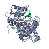 7yohC 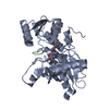 7yoiC  1y7lS S: Starting model for refinement C: citing same article ( |
|---|---|
| Similar structure data |
-Links
 PDBj
PDBj
- Assembly
| Deposited unit | 
| ||||||||
|---|---|---|---|---|---|---|---|---|---|
| 1 | 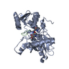
| ||||||||
| Unit cell |
|
-Components
| #1: Protein | / CSase / O-acetylserine (thiol)-lyase / OAS-TL / O-acetylserine sulfhydrylase / O-acetyl-L-serine sulfhydrylase Mass: 33707.527 Da / Num. of mol.: 1 / Mutation: D67E, A68P, L98I, A301S Source method: isolated from a genetically manipulated source Source: (gene. exp.) Haemophilus influenzae Rd KW20 (bacteria)Gene: cysK, HI_1103 / Production host: Escherichia coli BL21(DE3) (bacteria) / References: UniProt: P45040, cysteine synthase |
|---|---|
| #2: Protein/peptide | Mass: 900.930 Da / Num. of mol.: 1 / Source method: obtained synthetically / Source: (synth.) Salmonella typhimurium (bacteria) |
| #3: Water | ChemComp-HOH / Water Mass: 18.015 Da / Num. of mol.: 7 / Source method: isolated from a natural source / Formula: H2O Mass: 18.015 Da / Num. of mol.: 7 / Source method: isolated from a natural source / Formula: H2O |
| Has ligand of interest | Y |
-Experimental details
-Experiment
| Experiment | Method: X-RAY DIFFRACTION / Number of used crystals: 1 |
|---|
- Sample preparation
| Crystal | Density Matthews: 2.12 Å3/Da / Density % sol: 42.03 % |
|---|---|
| Crystal grow | Temperature: 278 K / Method: vapor diffusion, sitting drop / pH: 7.5 / Details: 0.1M HEPES, 1.3M Sodium citrate / PH range: 7.3-7.9 |
-Data collection
| Diffraction | Mean temperature: 100 K / Serial crystal experiment: N |
|---|---|
| Diffraction source | Source: ROTATING ANODE / Type: RIGAKU / Wavelength: 1.54179 Å |
| Detector | Type: MAR scanner 345 mm plate / Detector: IMAGE PLATE / Date: May 8, 2012 |
| Radiation | Monochromator: Mirrors / Protocol: SINGLE WAVELENGTH / Monochromatic (M) / Laue (L): M / Scattering type: x-ray |
| Radiation wavelength | Wavelength: 1.54179 Å / Relative weight: 1 |
| Reflection | Resolution: 2.8→40.831 Å / Num. obs: 69653 / % possible obs: 97.66 % / Redundancy: 10.1 % / Biso Wilson estimate: 24.22 Å2 / CC1/2: 0.713 / Rmerge(I) obs: 0.6911 / Net I/σ(I): 42.47 |
| Reflection shell | Resolution: 2.8→2.9 Å / Num. unique obs: 691 / CC1/2: 0.634 / CC star: 0.881 / % possible all: 100 |
- Processing
| Software |
| |||||||||||||||||||||||||||||||||||||||||||||||||||||||||||||||||||||||||||||||||||||||||||||||||||||||||||||||||||||||||||||||||||||||||||||||||||
|---|---|---|---|---|---|---|---|---|---|---|---|---|---|---|---|---|---|---|---|---|---|---|---|---|---|---|---|---|---|---|---|---|---|---|---|---|---|---|---|---|---|---|---|---|---|---|---|---|---|---|---|---|---|---|---|---|---|---|---|---|---|---|---|---|---|---|---|---|---|---|---|---|---|---|---|---|---|---|---|---|---|---|---|---|---|---|---|---|---|---|---|---|---|---|---|---|---|---|---|---|---|---|---|---|---|---|---|---|---|---|---|---|---|---|---|---|---|---|---|---|---|---|---|---|---|---|---|---|---|---|---|---|---|---|---|---|---|---|---|---|---|---|---|---|---|---|---|---|
| Refinement | Method to determine structure: MOLECULAR REPLACEMENT Starting model: 1Y7L Resolution: 2.8→40.831 Å / Cor.coef. Fo:Fc: 0.881 / Cor.coef. Fo:Fc free: 0.835 / SU B: 16.951 / SU ML: 0.339 / Cross valid method: FREE R-VALUE / ESU R Free: 0.467 Details: Hydrogens have been used if present in the input file
| |||||||||||||||||||||||||||||||||||||||||||||||||||||||||||||||||||||||||||||||||||||||||||||||||||||||||||||||||||||||||||||||||||||||||||||||||||
| Solvent computation | Ion probe radii: 0.8 Å / Shrinkage radii: 0.8 Å / VDW probe radii: 1.2 Å / Solvent model: MASK BULK SOLVENT | |||||||||||||||||||||||||||||||||||||||||||||||||||||||||||||||||||||||||||||||||||||||||||||||||||||||||||||||||||||||||||||||||||||||||||||||||||
| Displacement parameters | Biso mean: 22.582 Å2
| |||||||||||||||||||||||||||||||||||||||||||||||||||||||||||||||||||||||||||||||||||||||||||||||||||||||||||||||||||||||||||||||||||||||||||||||||||
| Refinement step | Cycle: LAST / Resolution: 2.8→40.831 Å
| |||||||||||||||||||||||||||||||||||||||||||||||||||||||||||||||||||||||||||||||||||||||||||||||||||||||||||||||||||||||||||||||||||||||||||||||||||
| Refine LS restraints |
| |||||||||||||||||||||||||||||||||||||||||||||||||||||||||||||||||||||||||||||||||||||||||||||||||||||||||||||||||||||||||||||||||||||||||||||||||||
| LS refinement shell |
|
