 Movie
Movie Controller


+ Open data

- Basic information
| Entry | Database: PDB / ID: 7ykn | ||||||
|---|---|---|---|---|---|---|---|
| Title | Crystal structure of (6-4) photolyase from Vibrio cholerae | ||||||
 Components Components | Cryptochrome/photolyase family protein | ||||||
 Keywords Keywords |  LYASE / Photolyase / DNA repair / DMRL / [4Fe-4S] cluster / FAD LYASE / Photolyase / DNA repair / DMRL / [4Fe-4S] cluster / FAD | ||||||
| Function / homology |  Function and homology information Function and homology information | ||||||
| Biological species |   Vibrio cholerae (bacteria) Vibrio cholerae (bacteria) | ||||||
| Method | X-RAY DIFFRACTION / MOLECULAR REPLACEMENT / Resolution: 2.5 Å | ||||||
 Authors Authors | Cakilkaya, B. / Kavakli, I.H. / DeMirci, H. | ||||||
| Funding support |  Turkey, 1items Turkey, 1items
| ||||||
 Citation Citation | Journal: J.Biol.Chem. / Year: 2023 Title: The crystal structure of Vibrio cholerae (6-4) photolyase reveals interactions with cofactors and a DNA-binding region. Authors: Cakilkaya, B. / Kavakli, I.H. / DeMirci, H. | ||||||
| History |
|
- Structure visualization
| Structure viewer | Molecule: MolmilJmol/JSmol |
|---|
- Downloads & links
-Download
| PDBx/mmCIF format | 7ykn.cif.gz | 238 KB | Display | PDBx/mmCIF format |
|---|---|---|---|---|
| PDB format | pdb7ykn.ent.gz | 185.9 KB | Display | PDB format |
| PDBx/mmJSON format | 7ykn.json.gz | Tree view | PDBx/mmJSON format | |
| Others |  Other downloads Other downloads |
-Validation report
| Arichive directory | https://data.pdbj.org/pub/pdb/validation_reports/yk/7yknftp://data.pdbj.org/pub/pdb/validation_reports/yk/7ykn | HTTPS FTP |
|---|
-Related structure data
| Related structure data | 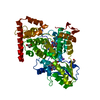 4djaS S: Starting model for refinement |
|---|---|
| Similar structure data |
-Links
 PDBj
PDBj


- Assembly
| Deposited unit | 
| ||||||||
|---|---|---|---|---|---|---|---|---|---|
| 1 |
| ||||||||
| Unit cell |
|
-Components
-Protein , 1 types, 1 molecules A
| #1: Protein | Mass: 61669.031 Da / Num. of mol.: 1 Source method: isolated from a genetically manipulated source Source: (gene. exp.) Vibrio cholerae (bacteria)Gene: D6U24_03430, ERS013165_01011, ERS013186_03677, ERS013198_00016, ERS013199_00375, ERS013202_01140, ERS013207_00426 Production host: Escherichia coli (E. coli) / References: UniProt: A0A2V4NVF4 |
|---|
-Non-polymers , 6 types, 563 molecules 


| #2: Chemical | Glycerol Mass: 92.094 Da / Num. of mol.: 2 / Source method: obtained synthetically / Formula: C3H8O3 Mass: 92.094 Da / Num. of mol.: 2 / Source method: obtained synthetically / Formula: C3H8O3#3: Chemical | ChemComp-DLZ / | 6,7-Dimethyl-8-ribityllumazine Mass: 326.305 Da / Num. of mol.: 1 / Source method: obtained synthetically / Formula: C13H18N4O6 / Feature type: SUBJECT OF INVESTIGATION Mass: 326.305 Da / Num. of mol.: 1 / Source method: obtained synthetically / Formula: C13H18N4O6 / Feature type: SUBJECT OF INVESTIGATION#4: Chemical | ChemComp-FAD / | Flavin adenine dinucleotide Mass: 785.550 Da / Num. of mol.: 1 / Source method: obtained synthetically / Formula: C27H33N9O15P2 / Feature type: SUBJECT OF INVESTIGATION / Comment: FAD*YM Mass: 785.550 Da / Num. of mol.: 1 / Source method: obtained synthetically / Formula: C27H33N9O15P2 / Feature type: SUBJECT OF INVESTIGATION / Comment: FAD*YM#5: Chemical | ChemComp-IMD / | Imidazole Mass: 69.085 Da / Num. of mol.: 1 / Source method: obtained synthetically / Formula: C3H5N2 Mass: 69.085 Da / Num. of mol.: 1 / Source method: obtained synthetically / Formula: C3H5N2#6: Chemical | ChemComp-SF4 / | Iron–sulfur cluster Mass: 351.640 Da / Num. of mol.: 1 / Source method: obtained synthetically / Formula: Fe4S4 / Feature type: SUBJECT OF INVESTIGATION Mass: 351.640 Da / Num. of mol.: 1 / Source method: obtained synthetically / Formula: Fe4S4 / Feature type: SUBJECT OF INVESTIGATION#7: Water | ChemComp-HOH / | WaterMass: 18.015 Da / Num. of mol.: 557 / Source method: isolated from a natural source / Formula: H2O |
|---|
-Details
| Has ligand of interest | Y |
|---|
-Experimental details
-Experiment
| Experiment | Method: X-RAY DIFFRACTION / Number of used crystals: 1 |
|---|
- Sample preparation
| Crystal | Density Matthews: 3.77 Å3/Da / Density % sol: 67.33 % |
|---|---|
| Crystal grow | Temperature: 277 K / Method: microbatch Details: 5%(v/v) PEG400, 2 M ammonium citrate/citric acid pH 7.5, 1 M sodium sulfate decahydrate |
-Data collection
| Diffraction | Mean temperature: 100 K / Serial crystal experiment: N |
|---|---|
| Diffraction source | Source: ROTATING ANODE / Type: RIGAKU PhotonJet-R / Wavelength: 1.54 Å |
| Detector | Type: RIGAKU HyPix-3000 / Detector: PIXEL / Date: Mar 10, 2022 |
| Radiation | Protocol: SINGLE WAVELENGTH / Monochromatic (M) / Laue (L): M / Scattering type: x-ray |
| Radiation wavelength | Wavelength: 1.54 Å / Relative weight: 1 |
| Reflection | Resolution: 2.5→24.62 Å / Num. obs: 32043 / % possible obs: 99.6 % / Redundancy: 60.4 % / Biso Wilson estimate: 15.97 Å2 / CC1/2: 0.955 / CC star: 0.988 / Rmerge(I) obs: 1.382 / Rpim(I) all: 0.1776 / Rrim(I) all: 1.394 / Net I/σ(I): 5.61 |
| Reflection shell | Resolution: 2.5→2.58 Å / Rmerge(I) obs: 3.765 / Mean I/σ(I) obs: 1.7 / Num. unique obs: 3130 / CC1/2: 0.546 / CC star: 0.84 / Rpim(I) all: 0.4855 / Rrim(I) all: 3.796 / % possible all: 99.97 |
- Processing
| Software |
| |||||||||||||||||||||||||||||||||||||||||||||||||||||||||||||||||||||||||||||||||||||||||||||||||||||||||||||||||||||||||||||||||||||||||||||||||||||||||||||||||||||||||||||||||||||||||||||||||||||||||||||||||||||||||||||||||
|---|---|---|---|---|---|---|---|---|---|---|---|---|---|---|---|---|---|---|---|---|---|---|---|---|---|---|---|---|---|---|---|---|---|---|---|---|---|---|---|---|---|---|---|---|---|---|---|---|---|---|---|---|---|---|---|---|---|---|---|---|---|---|---|---|---|---|---|---|---|---|---|---|---|---|---|---|---|---|---|---|---|---|---|---|---|---|---|---|---|---|---|---|---|---|---|---|---|---|---|---|---|---|---|---|---|---|---|---|---|---|---|---|---|---|---|---|---|---|---|---|---|---|---|---|---|---|---|---|---|---|---|---|---|---|---|---|---|---|---|---|---|---|---|---|---|---|---|---|---|---|---|---|---|---|---|---|---|---|---|---|---|---|---|---|---|---|---|---|---|---|---|---|---|---|---|---|---|---|---|---|---|---|---|---|---|---|---|---|---|---|---|---|---|---|---|---|---|---|---|---|---|---|---|---|---|---|---|---|---|---|---|---|---|---|---|---|---|---|---|---|---|---|---|---|---|---|
| Refinement | Method to determine structure: MOLECULAR REPLACEMENT Starting model: 4dja Resolution: 2.5→24.62 Å / SU ML: 0.32 / Cross valid method: THROUGHOUT / σ(F): 1.33 / Phase error: 25.02 / Stereochemistry target values: ML
| |||||||||||||||||||||||||||||||||||||||||||||||||||||||||||||||||||||||||||||||||||||||||||||||||||||||||||||||||||||||||||||||||||||||||||||||||||||||||||||||||||||||||||||||||||||||||||||||||||||||||||||||||||||||||||||||||
| Solvent computation | Shrinkage radii: 0.9 Å / VDW probe radii: 1.1 Å / Solvent model: FLAT BULK SOLVENT MODEL | |||||||||||||||||||||||||||||||||||||||||||||||||||||||||||||||||||||||||||||||||||||||||||||||||||||||||||||||||||||||||||||||||||||||||||||||||||||||||||||||||||||||||||||||||||||||||||||||||||||||||||||||||||||||||||||||||
| Displacement parameters | Biso max: 106.6 Å2 / Biso mean: 22.4624 Å2 / Biso min: 8.4 Å2 | |||||||||||||||||||||||||||||||||||||||||||||||||||||||||||||||||||||||||||||||||||||||||||||||||||||||||||||||||||||||||||||||||||||||||||||||||||||||||||||||||||||||||||||||||||||||||||||||||||||||||||||||||||||||||||||||||
| Refinement step | Cycle: final / Resolution: 2.5→24.62 Å
| |||||||||||||||||||||||||||||||||||||||||||||||||||||||||||||||||||||||||||||||||||||||||||||||||||||||||||||||||||||||||||||||||||||||||||||||||||||||||||||||||||||||||||||||||||||||||||||||||||||||||||||||||||||||||||||||||
| LS refinement shell | Refine-ID: X-RAY DIFFRACTION / Rfactor Rfree error: 0 / Total num. of bins used: 8
| |||||||||||||||||||||||||||||||||||||||||||||||||||||||||||||||||||||||||||||||||||||||||||||||||||||||||||||||||||||||||||||||||||||||||||||||||||||||||||||||||||||||||||||||||||||||||||||||||||||||||||||||||||||||||||||||||
| Refinement TLS params. | Method: refined / Refine-ID: X-RAY DIFFRACTION
| |||||||||||||||||||||||||||||||||||||||||||||||||||||||||||||||||||||||||||||||||||||||||||||||||||||||||||||||||||||||||||||||||||||||||||||||||||||||||||||||||||||||||||||||||||||||||||||||||||||||||||||||||||||||||||||||||
| Refinement TLS group |
|
