 Movie
Movie Controller


+ Open data

- Basic information
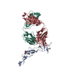
| Entry | Database: PDB / ID: 7yk4 | ||||||
|---|---|---|---|---|---|---|---|
| Title | ox40-antibody | ||||||
 Components Components |
| ||||||
 Keywords Keywords |  ANTITUMOR PROTEIN/IMMUNE SYSTEM / ox40 and antibody complex / ANTITUMOR PROTEIN / ANTITUMOR PROTEIN-IMMUNE SYSTEM complex ANTITUMOR PROTEIN/IMMUNE SYSTEM / ox40 and antibody complex / ANTITUMOR PROTEIN / ANTITUMOR PROTEIN-IMMUNE SYSTEM complex | ||||||
| Function / homology |  Function and homology informationtumor necrosis factor receptor activity / TNFs bind their physiological receptors / positive regulation of immunoglobulin production / T cell proliferation / positive regulation of B cell proliferation / negative regulation of DNA-binding transcription factor activity / virus receptor activity / inflammatory response / immune response / external side of plasma membrane ...tumor necrosis factor receptor activity / TNFs bind their physiological receptors / positive regulation of immunoglobulin production / T cell proliferation / positive regulation of B cell proliferation / negative regulation of DNA-binding transcription factor activity / virus receptor activity / inflammatory response / immune response / external side of plasma membrane / negative regulation of DNA-templated transcription / cell surface / plasma membrane Function and homology informationtumor necrosis factor receptor activity / TNFs bind their physiological receptors / positive regulation of immunoglobulin production / T cell proliferation / positive regulation of B cell proliferation / negative regulation of DNA-binding transcription factor activity / virus receptor activity / inflammatory response / immune response / external side of plasma membrane ...tumor necrosis factor receptor activity / TNFs bind their physiological receptors / positive regulation of immunoglobulin production / T cell proliferation / positive regulation of B cell proliferation / negative regulation of DNA-binding transcription factor activity / virus receptor activity / inflammatory response / immune response / external side of plasma membrane / negative regulation of DNA-templated transcription / cell surface / plasma membraneSimilarity search - Function | ||||||
| Biological species |  Homo sapiens (human) Homo sapiens (human) | ||||||
| Method | X-RAY DIFFRACTION / SYNCHROTRON / SAD / Resolution: 2.7 Å | ||||||
 Authors Authors | Zhou, A. | ||||||
| Funding support |  China, 1items China, 1items
| ||||||
 Citation Citation | Journal: Biomolecules / Year: 2022 Title: Structural Basis of a Novel Agonistic Anti-OX40 Antibody. Authors: Zhang, J. / Jiang, X. / Gao, H. / Zhang, F. / Zhang, X. / Zhou, A. / Xu, T. / Cai, H. | ||||||
| History |
|
- Structure visualization
| Structure viewer | Molecule: MolmilJmol/JSmol |
|---|
- Downloads & links
-Download
| PDBx/mmCIF format | 7yk4.cif.gz | 459.9 KB | Display | PDBx/mmCIF format |
|---|---|---|---|---|
| PDB format | pdb7yk4.ent.gz | 323.2 KB | Display | PDB format |
| PDBx/mmJSON format | 7yk4.json.gz | Tree view | PDBx/mmJSON format | |
| Others |  Other downloads Other downloads |
-Validation report
| Arichive directory | https://data.pdbj.org/pub/pdb/validation_reports/yk/7yk4ftp://data.pdbj.org/pub/pdb/validation_reports/yk/7yk4 | HTTPS FTP |
|---|
-Related structure data
-Links
 PDBj
PDBj


- Assembly
| Deposited unit | 
| ||||||||||||
|---|---|---|---|---|---|---|---|---|---|---|---|---|---|
| 1 | 
| ||||||||||||
| 2 | 
| ||||||||||||
| Unit cell |
|
-Components
| #1: Antibody | Mass: 25742.654 Da / Num. of mol.: 2 Source method: isolated from a genetically manipulated source Source: (gene. exp.) Homo sapiens (human) / Cell line (production host): HEK293 / Production host: Homo sapiens (human)#2: Antibody | Mass: 23531.135 Da / Num. of mol.: 2 Source method: isolated from a genetically manipulated source Source: (gene. exp.) Homo sapiens (human) / Cell line (production host): HEK293 / Production host: Homo sapiens (human)#3: Protein | Mass: 17027.002 Da / Num. of mol.: 2 Source method: isolated from a genetically manipulated source Source: (gene. exp.) Homo sapiens (human) / Gene: TNFRSF4, TXGP1L / Cell line (production host): HEK293 / Production host: Homo sapiens (human) / References: UniProt: P43489#4: Sugar | ChemComp-NAG / | N-Acetylglucosamine  Type: D-saccharide, beta linking / Mass: 221.208 Da / Num. of mol.: 1 / Source method: obtained synthetically / Formula: C8H15NO6 / Feature type: SUBJECT OF INVESTIGATION Type: D-saccharide, beta linking / Mass: 221.208 Da / Num. of mol.: 1 / Source method: obtained synthetically / Formula: C8H15NO6 / Feature type: SUBJECT OF INVESTIGATION#5: Water | ChemComp-HOH / | Water Mass: 18.015 Da / Num. of mol.: 1 / Source method: isolated from a natural source / Formula: H2O Mass: 18.015 Da / Num. of mol.: 1 / Source method: isolated from a natural source / Formula: H2OHas ligand of interest | Y | |
|---|
-Experimental details
-Experiment
| Experiment | Method: X-RAY DIFFRACTION / Number of used crystals: 1 |
|---|
- Sample preparation
| Crystal | Density Matthews: 2.86 Å3/Da / Density % sol: 57 % |
|---|---|
| Crystal grow | Temperature: 298 K / Method: vapor diffusion, sitting drop Details: 0.1 M potassium sodium tartrate, 14% PEG 3350, 10% glycerol |
-Data collection
| Diffraction | Mean temperature: 100 K / Serial crystal experiment: N |
|---|---|
| Diffraction source | Source: SYNCHROTRON / Site: SSRF / Beamline: BL17U1 / Wavelength: 0.9979 Å |
| Detector | Type: AGILENT ATLAS CCD / Detector: CCD / Date: Dec 21, 2017 |
| Radiation | Protocol: SINGLE WAVELENGTH / Monochromatic (M) / Laue (L): M / Scattering type: x-ray |
| Radiation wavelength | Wavelength: 0.9979 Å / Relative weight: 1 |
| Reflection | Resolution: 2.7→48.73 Å / Num. obs: 40452 / % possible obs: 94.2 % / Redundancy: 9.5 % / Biso Wilson estimate: 87.76 Å2 / CC1/2: 0.994 / Rmerge(I) obs: 0.154 / Net I/σ(I): 9.5 |
| Reflection shell | Resolution: 2.7→2.797 Å / Rmerge(I) obs: 0.992 / Num. unique obs: 40392 |
- Processing
| Software |
| ||||||||||||||||||||||||||||||||||||||||||||||||||||||||||||||||||||||||||||||||||||||||||||||||||||||||||||||||
|---|---|---|---|---|---|---|---|---|---|---|---|---|---|---|---|---|---|---|---|---|---|---|---|---|---|---|---|---|---|---|---|---|---|---|---|---|---|---|---|---|---|---|---|---|---|---|---|---|---|---|---|---|---|---|---|---|---|---|---|---|---|---|---|---|---|---|---|---|---|---|---|---|---|---|---|---|---|---|---|---|---|---|---|---|---|---|---|---|---|---|---|---|---|---|---|---|---|---|---|---|---|---|---|---|---|---|---|---|---|---|---|---|---|
| Refinement | Method to determine structure: SAD / Resolution: 2.7→48.73 Å / SU ML: 0.4542 / Cross valid method: FREE R-VALUE / σ(F): 1.38 / Phase error: 36.868 Stereochemistry target values: GeoStd + Monomer Library + CDL v1.2
| ||||||||||||||||||||||||||||||||||||||||||||||||||||||||||||||||||||||||||||||||||||||||||||||||||||||||||||||||
| Solvent computation | Shrinkage radii: 0.9 Å / VDW probe radii: 1.11 Å / Solvent model: FLAT BULK SOLVENT MODEL | ||||||||||||||||||||||||||||||||||||||||||||||||||||||||||||||||||||||||||||||||||||||||||||||||||||||||||||||||
| Displacement parameters | Biso mean: 114.26 Å2 | ||||||||||||||||||||||||||||||||||||||||||||||||||||||||||||||||||||||||||||||||||||||||||||||||||||||||||||||||
| Refinement step | Cycle: LAST / Resolution: 2.7→48.73 Å
| ||||||||||||||||||||||||||||||||||||||||||||||||||||||||||||||||||||||||||||||||||||||||||||||||||||||||||||||||
| Refine LS restraints |
| ||||||||||||||||||||||||||||||||||||||||||||||||||||||||||||||||||||||||||||||||||||||||||||||||||||||||||||||||
| LS refinement shell |
| ||||||||||||||||||||||||||||||||||||||||||||||||||||||||||||||||||||||||||||||||||||||||||||||||||||||||||||||||
| Refinement TLS params. | Method: refined / Origin x: -33.0256194596 Å / Origin y: -31.3828561399 Å / Origin z: -47.0764267477 Å
| ||||||||||||||||||||||||||||||||||||||||||||||||||||||||||||||||||||||||||||||||||||||||||||||||||||||||||||||||
| Refinement TLS group | Selection details: all |
