 Movie
Movie Controller


+ Open data

- Basic information
| Entry | Database: PDB / ID: 7yax | ||||||||||||
|---|---|---|---|---|---|---|---|---|---|---|---|---|---|
| Title | HYDROXYNITRILE LYASE FROM THE MILLIPEDE, | ||||||||||||
 Components Components | Hydroxynitrile lyase | ||||||||||||
 Keywords Keywords |  LYASE LYASE | ||||||||||||
| Function / homology | lyase activity / Hydroxynitrile lyase Function and homology information Function and homology information | ||||||||||||
| Biological species |  Oxidus gracilis (arthropod) Oxidus gracilis (arthropod) | ||||||||||||
| Method | X-RAY DIFFRACTION / MOLECULAR REPLACEMENT / Resolution: 2.01 Å | ||||||||||||
 Authors Authors | Chaikaew, S. / Watanabe, Y. / Zheng, D. / Motojima, F. / Asano, Y. | ||||||||||||
| Funding support |  Japan, 3items Japan, 3items
| ||||||||||||
 Citation Citation | Journal: To Be Published Title: Structure-based site-directed mutagenesis of hydroxynitrile lyase from cyanogenic millipede, Oxidus gracilis for enhancing catalytic efficiency and enantioselectivity Authors: Chaikaew, S. / Watanabe, Y. / Zheng, D. / Motojima, F. / Asano, Y. | ||||||||||||
| History |
|
- Structure visualization
| Structure viewer | Molecule: MolmilJmol/JSmol |
|---|
- Downloads & links
-Download
| PDBx/mmCIF format | 7yax.cif.gz | 274.9 KB | Display | PDBx/mmCIF format |
|---|---|---|---|---|
| PDB format | pdb7yax.ent.gz | 219.3 KB | Display | PDB format |
| PDBx/mmJSON format | 7yax.json.gz | Tree view | PDBx/mmJSON format | |
| Others |  Other downloads Other downloads |
-Validation report
| Arichive directory | https://data.pdbj.org/pub/pdb/validation_reports/ya/7yaxftp://data.pdbj.org/pub/pdb/validation_reports/ya/7yax | HTTPS FTP |
|---|
-Related structure data
| Related structure data |  7ycbC 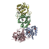 7ycdC 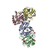 7ycfC 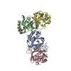 7yctC 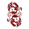 6kfeS S: Starting model for refinement C: citing same article ( |
|---|---|
| Similar structure data |
-Links
 PDBj
PDBj- Assembly
| Deposited unit | 
| ||||||||||||||||||
|---|---|---|---|---|---|---|---|---|---|---|---|---|---|---|---|---|---|---|---|
| 1 |
| ||||||||||||||||||
| Unit cell |
| ||||||||||||||||||
| Components on special symmetry positions |
|
-Components
| #1: Protein | Mass: 20383.896 Da / Num. of mol.: 4 Source method: isolated from a genetically manipulated source Source: (gene. exp.) Oxidus gracilis (arthropod) / Gene: OgraHNL / Production host:  Escherichia coli (E. coli) / References: UniProt: A0A2Z5XCT7 Escherichia coli (E. coli) / References: UniProt: A0A2Z5XCT7#2: Chemical | ChemComp-SO4 / Sulfate  Mass: 96.063 Da / Num. of mol.: 12 / Source method: obtained synthetically / Formula: SO4 / Feature type: SUBJECT OF INVESTIGATION Mass: 96.063 Da / Num. of mol.: 12 / Source method: obtained synthetically / Formula: SO4 / Feature type: SUBJECT OF INVESTIGATION#3: Chemical | ChemComp-CL / Chloride  Mass: 35.453 Da / Num. of mol.: 4 / Source method: isolated from a natural source / Formula: Cl / Feature type: SUBJECT OF INVESTIGATION Mass: 35.453 Da / Num. of mol.: 4 / Source method: isolated from a natural source / Formula: Cl / Feature type: SUBJECT OF INVESTIGATION#4: Water | ChemComp-HOH / | Water Mass: 18.015 Da / Num. of mol.: 865 / Source method: isolated from a natural source / Formula: H2O Mass: 18.015 Da / Num. of mol.: 865 / Source method: isolated from a natural source / Formula: H2OHas ligand of interest | Y | |
|---|
-Experimental details
-Experiment
| Experiment | Method: X-RAY DIFFRACTION / Number of used crystals: 1 |
|---|
- Sample preparation
| Crystal | Density Matthews: 3.49 Å3/Da / Density % sol: 68.8 % |
|---|---|
| Crystal grow | Temperature: 293.15 K / Method: vapor diffusion, sitting drop / Details: 0.1 M BIS-TRIS (pH 5.5), 2.0 M ammonium sulfate |
-Data collection
| Diffraction | Mean temperature: 100 K / Serial crystal experiment: N |
|---|---|
| Diffraction source | Source: ROTATING ANODE / Type: RIGAKU / Wavelength: 1.5418 Å |
| Detector | Type: RIGAKU / Detector: IMAGE PLATE / Date: Jul 5, 2017 |
| Radiation | Protocol: SINGLE WAVELENGTH / Monochromatic (M) / Laue (L): M / Scattering type: x-ray |
| Radiation wavelength | Wavelength: 1.5418 Å / Relative weight: 1 |
| Reflection | Resolution: 2.01→64.57 Å / Num. obs: 595005 / % possible obs: 99.58 % / Redundancy: 8 % / Rmerge(I) obs: 0.061 / Net I/σ(I): 20.6 |
| Reflection shell | Resolution: 2.01→2.05 Å / Rmerge(I) obs: 0.221 / Num. unique obs: 4547 |
- Processing
| Software |
| |||||||||||||||||||||||||
|---|---|---|---|---|---|---|---|---|---|---|---|---|---|---|---|---|---|---|---|---|---|---|---|---|---|---|
| Refinement | Method to determine structure: MOLECULAR REPLACEMENT Starting model: 6KFE Resolution: 2.01→39.969 Å / Cor.coef. Fo:Fc: 0.963 / Cor.coef. Fo:Fc free: 0.946 / SU B: 2.225 / SU ML: 0.063 / Cross valid method: THROUGHOUT / ESU R: 0.108 / ESU R Free: 0.108 Details: Hydrogens have been added in their riding positions
| |||||||||||||||||||||||||
| Solvent computation | Ion probe radii: 0.8 Å / Shrinkage radii: 0.8 Å / VDW probe radii: 1.2 Å / Solvent model: MASK BULK SOLVENT | |||||||||||||||||||||||||
| Displacement parameters | Biso mean: 19.132 Å2
| |||||||||||||||||||||||||
| Refinement step | Cycle: LAST / Resolution: 2.01→39.969 Å
| |||||||||||||||||||||||||
| Refine LS restraints | Type: r_lrange_other / Dev ideal: 6.104 / Dev ideal target: 27.828 / Number: 6049 | |||||||||||||||||||||||||
| LS refinement shell | Resolution: 6.279→8.772 Å
|
