 Movie
Movie Controller


+ Open data

- Basic information
| Entry | Database: PDB / ID: 7y74 | ||||||
|---|---|---|---|---|---|---|---|
| Title | Apostichopus japonicus ferritin mutant-D129A/E132A | ||||||
 Components Components | Ferritin | ||||||
 Keywords Keywords | METAL BINDING PROTEIN / ion binding / ferroxidase activity | ||||||
| Function / homology |  Function and homology informationferroxidase / ferroxidase activity / ferric iron binding / iron ion transport / intracellular iron ion homeostasis Function and homology informationferroxidase / ferroxidase activity / ferric iron binding / iron ion transport / intracellular iron ion homeostasisSimilarity search - Function | ||||||
| Biological species |  Apostichopus japonicus (Japanese sea cucumber) Apostichopus japonicus (Japanese sea cucumber) | ||||||
| Method | X-RAY DIFFRACTION / SYNCHROTRON / MOLECULAR REPLACEMENT / Resolution: 1.98 Å | ||||||
 Authors Authors | Wu, Y. / Ming, T.H. / Su, X.R. | ||||||
| Funding support |  China, 1items China, 1items
| ||||||
 Citation Citation | Journal: To Be Published Title: Apostichopus japonicus ferritin mutant-D129A/E132A Authors: Wu, Y. | ||||||
| History |
|
- Structure visualization
| Structure viewer | Molecule: MolmilJmol/JSmol |
|---|
- Downloads & links
-Download
| PDBx/mmCIF format | 7y74.cif.gz | 442 KB | Display | PDBx/mmCIF format |
|---|---|---|---|---|
| PDB format | pdb7y74.ent.gz | 358.8 KB | Display | PDB format |
| PDBx/mmJSON format | 7y74.json.gz | Tree view | PDBx/mmJSON format | |
| Others |  Other downloads Other downloads |
-Validation report
| Arichive directory | https://data.pdbj.org/pub/pdb/validation_reports/y7/7y74ftp://data.pdbj.org/pub/pdb/validation_reports/y7/7y74 | HTTPS FTP |
|---|
-Related structure data
| Related structure data | 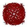 5wpnS S: Starting model for refinement |
|---|---|
| Similar structure data |
-Links
 PDBj
PDBj


- Assembly
| Deposited unit | 
| ||||||||
|---|---|---|---|---|---|---|---|---|---|
| 1 | 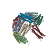
| ||||||||
| Unit cell |
| ||||||||
| Components on special symmetry positions |
|
-Components
| #1: Protein | Mass: 19966.545 Da / Num. of mol.: 12 / Mutation: D129A,E132A Source method: isolated from a genetically manipulated source Source: (gene. exp.) Apostichopus japonicus (Japanese sea cucumber)Gene: BSL78_19566 / Production host:  Escherichia coli (E. coli) / References: UniProt: Q2QJV4, ferroxidase Escherichia coli (E. coli) / References: UniProt: Q2QJV4, ferroxidase#2: Chemical | ChemComp-FE / Iron  Mass: 55.845 Da / Num. of mol.: 36 / Source method: obtained synthetically / Formula: Fe / Feature type: SUBJECT OF INVESTIGATION Mass: 55.845 Da / Num. of mol.: 36 / Source method: obtained synthetically / Formula: Fe / Feature type: SUBJECT OF INVESTIGATION#3: Chemical | ChemComp-CD /   Mass: 112.411 Da / Num. of mol.: 12 / Source method: isolated from a natural source / Formula: Cd Mass: 112.411 Da / Num. of mol.: 12 / Source method: isolated from a natural source / Formula: Cd#4: Water | ChemComp-HOH / | Water Mass: 18.015 Da / Num. of mol.: 1636 / Source method: isolated from a natural source / Formula: H2O Mass: 18.015 Da / Num. of mol.: 1636 / Source method: isolated from a natural source / Formula: H2OHas ligand of interest | Y | |
|---|
-Experimental details
-Experiment
| Experiment | Method: X-RAY DIFFRACTION / Number of used crystals: 1 |
|---|
- Sample preparation
| Crystal | Density Matthews: 2.81 Å3/Da / Density % sol: 56.3 % |
|---|---|
| Crystal grow | Temperature: 289 K / Method: vapor diffusion, sitting drop Details: 0.05 M Cadmium sulfate hydrate, 0.1 M HEPES pH 7.5, 1.0 M Sodium acetate trihydrate |
-Data collection
| Diffraction | Mean temperature: 100 K / Serial crystal experiment: N | |||||||||||||||||||||||||||||||||||||||||||||||||||||||||||||||||||||||||||||||||||||||||||||||||||||||||||||||||||||||||||||||||||||||||||||||||||||||||||||||||||||||||||||||||||||||||||||
|---|---|---|---|---|---|---|---|---|---|---|---|---|---|---|---|---|---|---|---|---|---|---|---|---|---|---|---|---|---|---|---|---|---|---|---|---|---|---|---|---|---|---|---|---|---|---|---|---|---|---|---|---|---|---|---|---|---|---|---|---|---|---|---|---|---|---|---|---|---|---|---|---|---|---|---|---|---|---|---|---|---|---|---|---|---|---|---|---|---|---|---|---|---|---|---|---|---|---|---|---|---|---|---|---|---|---|---|---|---|---|---|---|---|---|---|---|---|---|---|---|---|---|---|---|---|---|---|---|---|---|---|---|---|---|---|---|---|---|---|---|---|---|---|---|---|---|---|---|---|---|---|---|---|---|---|---|---|---|---|---|---|---|---|---|---|---|---|---|---|---|---|---|---|---|---|---|---|---|---|---|---|---|---|---|---|---|---|---|---|---|
| Diffraction source | Source: SYNCHROTRON / Site: SSRF / Beamline: BL19U1 / Wavelength: 0.97892 Å | |||||||||||||||||||||||||||||||||||||||||||||||||||||||||||||||||||||||||||||||||||||||||||||||||||||||||||||||||||||||||||||||||||||||||||||||||||||||||||||||||||||||||||||||||||||||||||||
| Detector | Type: DECTRIS PILATUS3 6M / Detector: PIXEL / Date: Jul 5, 2018 | |||||||||||||||||||||||||||||||||||||||||||||||||||||||||||||||||||||||||||||||||||||||||||||||||||||||||||||||||||||||||||||||||||||||||||||||||||||||||||||||||||||||||||||||||||||||||||||
| Radiation | Protocol: SINGLE WAVELENGTH / Monochromatic (M) / Laue (L): M / Scattering type: x-ray | |||||||||||||||||||||||||||||||||||||||||||||||||||||||||||||||||||||||||||||||||||||||||||||||||||||||||||||||||||||||||||||||||||||||||||||||||||||||||||||||||||||||||||||||||||||||||||||
| Radiation wavelength | Wavelength: 0.97892 Å / Relative weight: 1 | |||||||||||||||||||||||||||||||||||||||||||||||||||||||||||||||||||||||||||||||||||||||||||||||||||||||||||||||||||||||||||||||||||||||||||||||||||||||||||||||||||||||||||||||||||||||||||||
| Reflection | Resolution: 1.98→50 Å / Num. obs: 182173 / % possible obs: 99 % / Redundancy: 3.4 % / Rmerge(I) obs: 0.054 / Rpim(I) all: 0.034 / Rrim(I) all: 0.064 / Χ2: 0.864 / Net I/σ(I): 8.6 / Num. measured all: 617526 | |||||||||||||||||||||||||||||||||||||||||||||||||||||||||||||||||||||||||||||||||||||||||||||||||||||||||||||||||||||||||||||||||||||||||||||||||||||||||||||||||||||||||||||||||||||||||||||
| Reflection shell | Diffraction-ID: 1
|
- Processing
| Software |
| ||||||||||||||||||||||||||||||||||||||||||||||||||||||||||||
|---|---|---|---|---|---|---|---|---|---|---|---|---|---|---|---|---|---|---|---|---|---|---|---|---|---|---|---|---|---|---|---|---|---|---|---|---|---|---|---|---|---|---|---|---|---|---|---|---|---|---|---|---|---|---|---|---|---|---|---|---|---|
| Refinement | Method to determine structure: MOLECULAR REPLACEMENT Starting model: 5WPN Resolution: 1.98→47.91 Å / Cor.coef. Fo:Fc: 0.953 / Cor.coef. Fo:Fc free: 0.934 / SU B: 3.326 / SU ML: 0.094 / Cross valid method: THROUGHOUT / σ(F): 0 / ESU R: 0.152 / ESU R Free: 0.144 / Stereochemistry target values: MAXIMUM LIKELIHOOD Details: HYDROGENS HAVE BEEN ADDED IN THE RIDING POSITIONS U VALUES : REFINED INDIVIDUALLY
| ||||||||||||||||||||||||||||||||||||||||||||||||||||||||||||
| Solvent computation | Ion probe radii: 0.8 Å / Shrinkage radii: 0.8 Å / VDW probe radii: 1.2 Å / Solvent model: MASK | ||||||||||||||||||||||||||||||||||||||||||||||||||||||||||||
| Displacement parameters | Biso max: 88.36 Å2 / Biso mean: 27.793 Å2 / Biso min: 12.34 Å2
| ||||||||||||||||||||||||||||||||||||||||||||||||||||||||||||
| Refinement step | Cycle: final / Resolution: 1.98→47.91 Å
| ||||||||||||||||||||||||||||||||||||||||||||||||||||||||||||
| Refine LS restraints |
| ||||||||||||||||||||||||||||||||||||||||||||||||||||||||||||
| LS refinement shell | Resolution: 1.981→2.032 Å / Rfactor Rfree error: 0 / Total num. of bins used: 20
|
