 Movie
Movie Controller


+ Open data

- Basic information
| Entry | Database: PDB / ID: 7xyt | ||||||||||||
|---|---|---|---|---|---|---|---|---|---|---|---|---|---|
| Title | Crystal structure of ZER1 bound to AFLH degron | ||||||||||||
 Components Components | Protein zer-1 homolog | ||||||||||||
 Keywords Keywords |  LIGASE / E3 LIGASE / E3 | ||||||||||||
| Function / homology | Cul2-RING ubiquitin ligase complex / protein quality control for misfolded or incompletely synthesized proteins / Armadillo/beta-catenin-like repeats / Armadillo / Leucine-rich repeat domain superfamily / positive regulation of proteasomal ubiquitin-dependent protein catabolic process / Armadillo-like helical / Armadillo-type fold / Protein zer-1 homolog Function and homology information Function and homology information | ||||||||||||
| Biological species |  Homo sapiens (human) Homo sapiens (human) | ||||||||||||
| Method | X-RAY DIFFRACTION / SYNCHROTRON / MOLECULAR REPLACEMENT / Resolution: 1.5 Å | ||||||||||||
 Authors Authors | Dong, C. / Yan, X. / Li, Y. | ||||||||||||
| Funding support |  China, 3items China, 3items
| ||||||||||||
 Citation Citation | Journal: Nat Commun / Year: 2022 Title: CRL2 ZER1/ZYG11B recognizes small N-terminal residues for degradation. Authors: Li, Y. / Zhao, Y. / Yan, X. / Ye, C. / Weirich, S. / Zhang, B. / Wang, X. / Song, L. / Jiang, C. / Jeltsch, A. / Dong, C. / Mi, W. #1: Journal: Nat Commun / Year: 2022Title: CRL2ZER1/ZYG11B recognizes small N-terminal residues for degradation Authors: Li, Y. / Zhao, Y. / Yan, X. / Ye, C. / Weirich, S. / Zhang, B. / Wang, X. / Song, L. / Jiang, C. / Jeltsch, A. / Dong, C. / Mi, W. | ||||||||||||
| History |
|
- Structure visualization
| Structure viewer | Molecule: MolmilJmol/JSmol |
|---|
- Downloads & links
-Download
| PDBx/mmCIF format | 7xyt.cif.gz | 439.5 KB | Display | PDBx/mmCIF format |
|---|---|---|---|---|
| PDB format | pdb7xyt.ent.gz | 362 KB | Display | PDB format |
| PDBx/mmJSON format | 7xyt.json.gz | Tree view | PDBx/mmJSON format | |
| Others |  Other downloads Other downloads |
-Validation report
| Arichive directory | https://data.pdbj.org/pub/pdb/validation_reports/xy/7xytftp://data.pdbj.org/pub/pdb/validation_reports/xy/7xyt | HTTPS FTP |
|---|
-Related structure data
| Related structure data |  7xysC 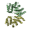 7xyuC 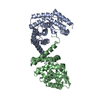 7xyvC 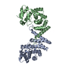 7xywC 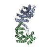 7xyxC  7ep3S S: Starting model for refinement C: citing same article ( |
|---|---|
| Similar structure data |
-Links
 PDBj
PDBj


- Assembly
| Deposited unit | 
| ||||||||
|---|---|---|---|---|---|---|---|---|---|
| 1 | 
| ||||||||
| 2 | 
| ||||||||
| Unit cell |
|
-Components
| #1: Protein | Mass: 29308.936 Da / Num. of mol.: 4 Source method: isolated from a genetically manipulated source Source: (gene. exp.) Homo sapiens (human) / Gene: ZER1, C9orf60, ZYG, ZYG11BL / Production host:  Escherichia coli (E. coli) / References: UniProt: Q7Z7L7 Escherichia coli (E. coli) / References: UniProt: Q7Z7L7#2: Water | ChemComp-HOH / | Water Mass: 18.015 Da / Num. of mol.: 1059 / Source method: isolated from a natural source / Formula: H2O Mass: 18.015 Da / Num. of mol.: 1059 / Source method: isolated from a natural source / Formula: H2O |
|---|
-Experimental details
-Experiment
| Experiment | Method: X-RAY DIFFRACTION / Number of used crystals: 1 |
|---|
- Sample preparation
| Crystal | Density Matthews: 2.33 Å3/Da / Density % sol: 47.18 % |
|---|---|
| Crystal grow | Temperature: 291 K / Method: vapor diffusion, sitting drop / Details: 0.2M NH4F, 20% PEG3350 |
-Data collection
| Diffraction | Mean temperature: 100 K / Serial crystal experiment: N |
|---|---|
| Diffraction source | Source: SYNCHROTRON / Site: SSRF / Beamline: BL02U1 / Wavelength: 0.9792 Å |
| Detector | Type: DECTRIS EIGER2 S 9M / Detector: PIXEL / Date: Jul 3, 2021 |
| Radiation | Protocol: SINGLE WAVELENGTH / Monochromatic (M) / Laue (L): M / Scattering type: x-ray |
| Radiation wavelength | Wavelength: 0.9792 Å / Relative weight: 1 |
| Reflection | Resolution: 1.5→138.27 Å / Num. obs: 170861 / % possible obs: 99.9 % / Redundancy: 6.6 % / Rmerge(I) obs: 0.118 / Net I/σ(I): 8.97 |
| Reflection shell | Resolution: 1.5→1.54 Å / Rmerge(I) obs: 1.568 / Num. unique obs: 12605 |
- Processing
| Software |
| |||||||||||||||||||||||||||||||||||||||||||||||||||||||||||||||||||||||||||||||||||||||||||||||||||||||||||||||||||||||||||||||||||||||||||||||||||||||||||||||||||||||||||||||||||||||||||||||||||||||||||||||||||||||||
|---|---|---|---|---|---|---|---|---|---|---|---|---|---|---|---|---|---|---|---|---|---|---|---|---|---|---|---|---|---|---|---|---|---|---|---|---|---|---|---|---|---|---|---|---|---|---|---|---|---|---|---|---|---|---|---|---|---|---|---|---|---|---|---|---|---|---|---|---|---|---|---|---|---|---|---|---|---|---|---|---|---|---|---|---|---|---|---|---|---|---|---|---|---|---|---|---|---|---|---|---|---|---|---|---|---|---|---|---|---|---|---|---|---|---|---|---|---|---|---|---|---|---|---|---|---|---|---|---|---|---|---|---|---|---|---|---|---|---|---|---|---|---|---|---|---|---|---|---|---|---|---|---|---|---|---|---|---|---|---|---|---|---|---|---|---|---|---|---|---|---|---|---|---|---|---|---|---|---|---|---|---|---|---|---|---|---|---|---|---|---|---|---|---|---|---|---|---|---|---|---|---|---|---|---|---|---|---|---|---|---|---|---|---|---|---|---|---|---|
| Refinement | Method to determine structure: MOLECULAR REPLACEMENT Starting model: 7EP3 Resolution: 1.5→34.61 Å / SU ML: 0.17 / Cross valid method: THROUGHOUT / σ(F): 1.34 / Phase error: 22.49 / Stereochemistry target values: ML
| |||||||||||||||||||||||||||||||||||||||||||||||||||||||||||||||||||||||||||||||||||||||||||||||||||||||||||||||||||||||||||||||||||||||||||||||||||||||||||||||||||||||||||||||||||||||||||||||||||||||||||||||||||||||||
| Solvent computation | Shrinkage radii: 0.9 Å / VDW probe radii: 1.11 Å / Solvent model: FLAT BULK SOLVENT MODEL | |||||||||||||||||||||||||||||||||||||||||||||||||||||||||||||||||||||||||||||||||||||||||||||||||||||||||||||||||||||||||||||||||||||||||||||||||||||||||||||||||||||||||||||||||||||||||||||||||||||||||||||||||||||||||
| Refinement step | Cycle: LAST / Resolution: 1.5→34.61 Å
| |||||||||||||||||||||||||||||||||||||||||||||||||||||||||||||||||||||||||||||||||||||||||||||||||||||||||||||||||||||||||||||||||||||||||||||||||||||||||||||||||||||||||||||||||||||||||||||||||||||||||||||||||||||||||
| Refine LS restraints |
| |||||||||||||||||||||||||||||||||||||||||||||||||||||||||||||||||||||||||||||||||||||||||||||||||||||||||||||||||||||||||||||||||||||||||||||||||||||||||||||||||||||||||||||||||||||||||||||||||||||||||||||||||||||||||
| LS refinement shell |
| |||||||||||||||||||||||||||||||||||||||||||||||||||||||||||||||||||||||||||||||||||||||||||||||||||||||||||||||||||||||||||||||||||||||||||||||||||||||||||||||||||||||||||||||||||||||||||||||||||||||||||||||||||||||||
| Refinement TLS params. | Method: refined / Origin x: 16.0207 Å / Origin y: 13.2666 Å / Origin z: 0.7674 Å
| |||||||||||||||||||||||||||||||||||||||||||||||||||||||||||||||||||||||||||||||||||||||||||||||||||||||||||||||||||||||||||||||||||||||||||||||||||||||||||||||||||||||||||||||||||||||||||||||||||||||||||||||||||||||||
| Refinement TLS group | Selection details: all |
