Journal: Nature / Year: 2023 Title: Structure of the human DICER-pre-miRNA complex in a dicing state. Authors: Young-Yoon Lee / Hansol Lee / Haedong Kim / V Narry Kim / Soung-Hun Roh / Abstract: Dicer has a key role in small RNA biogenesis, processing double-stranded RNAs (dsRNAs). Human DICER (hDICER, also known as DICER1) is specialized for cleaving small hairpin structures such as ...Dicer has a key role in small RNA biogenesis, processing double-stranded RNAs (dsRNAs). Human DICER (hDICER, also known as DICER1) is specialized for cleaving small hairpin structures such as precursor microRNAs (pre-miRNAs) and has limited activity towards long dsRNAs-unlike its homologues in lower eukaryotes and plants, which cleave long dsRNAs. Although the mechanism by which long dsRNAs are cleaved has been well documented, our understanding of pre-miRNA processing is incomplete because structures of hDICER in a catalytic state are lacking. Here we report the cryo-electron microscopy structure of hDICER bound to pre-miRNA in a dicing state and uncover the structural basis of pre-miRNA processing. hDICER undergoes large conformational changes to attain the active state. The helicase domain becomes flexible, which allows the binding of pre-miRNA to the catalytic valley. The double-stranded RNA-binding domain relocates and anchors pre-miRNA in a specific position through both sequence-independent and sequence-specific recognition of the newly identified 'GYM motif'. The DICER-specific PAZ helix is also reoriented to accommodate the RNA. Furthermore, our structure identifies a configuration of the 5' end of pre-miRNA inserted into a basic pocket. In this pocket, a group of arginine residues recognize the 5' terminal base (disfavouring guanine) and terminal monophosphate; this explains the specificity of hDICER and how it determines the cleavage site. We identify cancer-associated mutations in the 5' pocket residues that impair miRNA biogenesis. Our study reveals how hDICER recognizes pre-miRNAs with stringent specificity and enables a mechanistic understanding of hDICER-related diseases.
EndoribonucleaseDicer / Dicer / Helicase with RNase motif / Helicase MOI
Mass: 218947.328 Da / Num. of mol.: 1 Source method: isolated from a genetically manipulated source Source: (gene. exp.) Homo sapiens (human) / Gene: DICER1, DICER, HERNA, KIAA0928 / Production host: Homo sapiens (human) / References: UniProt: Q9UPY3, ribonuclease III
-
Experimental details
-
Experiment
Experiment
Method: ELECTRON MICROSCOPY
EM experiment
Aggregation state: 3D ARRAY / 3D reconstruction method: single particle reconstruction
-
Sample preparation
Component
Name: monomeric form of human DICER / Type: COMPLEX / Entity ID: all / Source: RECOMBINANT
Source (natural)
Organism: Homo sapiens (human)
Source (recombinant)
Organism: Homo sapiens (human)
Buffer solution
pH: 8
Specimen
Embedding applied: NO / Shadowing applied: NO / Staining applied: NO / Vitrification applied: YES
In the structure databanks used in Yorodumi, some data are registered as the other names, "COVID-19 virus" and "2019-nCoV". Here are the details of the virus and the list of structure data.
Jan 31, 2019. EMDB accession codes are about to change! (news from PDBe EMDB page)
EMDB accession codes are about to change! (news from PDBe EMDB page)
The allocation of 4 digits for EMDB accession codes will soon come to an end. Whilst these codes will remain in use, new EMDB accession codes will include an additional digit and will expand incrementally as the available range of codes is exhausted. The current 4-digit format prefixed with “EMD-” (i.e. EMD-XXXX) will advance to a 5-digit format (i.e. EMD-XXXXX), and so on. It is currently estimated that the 4-digit codes will be depleted around Spring 2019, at which point the 5-digit format will come into force.
The EM Navigator/Yorodumi systems omit the EMD- prefix.
Related info.:Q: What is EMD? / ID/Accession-code notation in Yorodumi/EM Navigator
Yorodumi is a browser for structure data from EMDB, PDB, SASBDB, etc.
This page is also the successor to EM Navigator detail page, and also detail information page/front-end page for Omokage search.
The word "yorodu" (or yorozu) is an old Japanese word meaning "ten thousand". "mi" (miru) is to see.
Related info.:EMDB / PDB / SASBDB / Comparison of 3 databanks / Yorodumi Search / Aug 31, 2016. New EM Navigator & Yorodumi / Yorodumi Papers / Jmol/JSmol / Function and homology information / Changes in new EM Navigator and Yorodumi
 Movie
Movie Controller


 Open data
 Basic information
 Components
 Dicer
Dicer  Keywords
 Function and homology information
 Authors
 Korea, Republic Of, 1items
  Citation
 Structure visualization
 Downloads & links
 Other downloads


 PDBj
PDBj


 Assembly
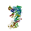
 Sample preparation
 Electron microscopy imaging
 Processing