 Movie
Movie Controller

[English] 日本語
 Yorodumi
- PDB-7xpe: Cryo-EM structure of the T=4 lake sinai virus 2 virus-like capsid... -
+ Open data

- Basic information
| Entry | Database: PDB / ID: 7xpe | ||||||
|---|---|---|---|---|---|---|---|
| Title | Cryo-EM structure of the T=4 lake sinai virus 2 virus-like capsid at pH 8.5 | ||||||
 Components Components | Capsid protein alpha | ||||||
 Keywords Keywords | VIRUS LIKE PARTICLE | ||||||
| Function / homology | Viral coat protein subunit / Capsid protein alpha Function and homology information Function and homology information | ||||||
| Biological species |  Lake Sinai virus 2 Lake Sinai virus 2 | ||||||
| Method | ELECTRON MICROSCOPY / single particle reconstruction / cryo EM / Resolution: 3.32 Å | ||||||
 Authors Authors | Chen, N.C. / Wang, C.H. / Chen, C.J. / Yoshimura, M. / Guan, H.H. / Chuankhayan, P. / Lin, C.C. | ||||||
| Funding support |  Taiwan, 1items Taiwan, 1items
| ||||||
 Citation Citation | Journal: Nat Commun / Year: 2023 Title: Structures of honeybee-infecting Lake Sinai virus reveal domain functions and capsid assembly with dynamic motions. Authors: Nai-Chi Chen / Chun-Hsiung Wang / Masato Yoshimura / Yi-Qi Yeh / Hong-Hsiang Guan / Phimonphan Chuankhayan / Chien-Chih Lin / Pei-Ju Lin / Yen-Chieh Huang / Soichi Wakatsuki / Meng-Chiao Ho / Chun-Jung Chen /   Abstract: Understanding the structural diversity of honeybee-infecting viruses is critical to maintain pollinator health and manage the spread of diseases in ecology and agriculture. We determine cryo-EM ...Understanding the structural diversity of honeybee-infecting viruses is critical to maintain pollinator health and manage the spread of diseases in ecology and agriculture. We determine cryo-EM structures of T = 4 and T = 3 capsids of virus-like particles (VLPs) of Lake Sinai virus (LSV) 2 and delta-N48 LSV1, belonging to tetraviruses, at resolutions of 2.3-2.6 Å in various pH environments. Structural analysis shows that the LSV2 capsid protein (CP) structural features, particularly the protruding domain and C-arm, differ from those of other tetraviruses. The anchor loop on the central β-barrel domain interacts with the neighboring subunit to stabilize homo-trimeric capsomeres during assembly. Delta-N48 LSV1 CP interacts with ssRNA via the rigid helix α1', α1'-α1 loop, β-barrel domain, and C-arm. Cryo-EM reconstructions, combined with X-ray crystallographic and small-angle scattering analyses, indicate that pH affects capsid conformations by regulating reversible dynamic particle motions and sizes of LSV2 VLPs. C-arms exist in all LSV2 and delta-N48 LSV1 VLPs across varied pH conditions, indicating that autoproteolysis cleavage is not required for LSV maturation. The observed linear domino-scaffold structures of various lengths, made up of trapezoid-shape capsomeres, provide a basis for icosahedral T = 4 and T = 3 architecture assemblies. These findings advance understanding of honeybee-infecting viruses that can cause Colony Collapse Disorder. | ||||||
| History |
|
- Structure visualization
| Structure viewer | Molecule: MolmilJmol/JSmol |
|---|
- Downloads & links
-Download
| PDBx/mmCIF format | 7xpe.cif.gz | 309.6 KB | Display | PDBx/mmCIF format |
|---|---|---|---|---|
| PDB format | pdb7xpe.ent.gz | 255.3 KB | Display | PDB format |
| PDBx/mmJSON format | 7xpe.json.gz | Tree view | PDBx/mmJSON format | |
| Others |  Other downloads Other downloads |
-Validation report
| Arichive directory | https://data.pdbj.org/pub/pdb/validation_reports/xp/7xpeftp://data.pdbj.org/pub/pdb/validation_reports/xp/7xpe | HTTPS FTP |
|---|
-Related structure data
| Related structure data |  33371MC  7xpaC  7xpbC 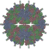 7xpdC  7xpfC  7xpgC M: map data used to model this data C: citing same article ( |
|---|---|
| Similar structure data |


-Links
 PDBj
PDBj- Assembly
| Deposited unit | 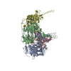
|
|---|---|
| 1 | x 60
|
| 2 |
|
| 3 | x 5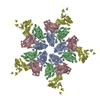
|
| 4 | x 6
|
| 5 | 
|
| Symmetry | Point symmetry: (Schoenflies symbol: I (icosahedral)) |
-Components
| #1: Protein | Mass: 57344.309 Da / Num. of mol.: 4 Source method: isolated from a genetically manipulated source Source: (gene. exp.) Lake Sinai virus 2 / Production host:  Escherichia coli (E. coli) / References: UniProt: F8TEV4 Escherichia coli (E. coli) / References: UniProt: F8TEV4 |
|---|
-Experimental details
-Experiment
| Experiment | Method: ELECTRON MICROSCOPY |
|---|---|
| EM experiment | Aggregation state: PARTICLE / 3D reconstruction method: single particle reconstruction |
- Sample preparation
| Component | Name: Lake Sinai virus 2 / Type: VIRUS / Entity ID: all / Source: RECOMBINANT |
|---|---|
| Source (natural) | Organism: Lake Sinai virus 2 |
| Source (recombinant) | Organism: Escherichia coli (E. coli) |
| Details of virus | Empty: YES / Enveloped: NO / Isolate: STRAIN / Type: VIRUS-LIKE PARTICLE |
| Buffer solution | pH: 7.5 |
| Specimen | Embedding applied: NO / Shadowing applied: NO / Staining applied: NO / Vitrification applied: YES |
| Vitrification | Instrument: FEI VITROBOT MARK IV / Cryogen name: ETHANE / Humidity: 100 % |
- Electron microscopy imaging
| Experimental equipment |  Model: Titan Krios / Image courtesy: FEI Company |
|---|---|
| Microscopy | Model: FEI TITAN KRIOS |
| Electron gun | Electron source: FIELD EMISSION GUN / Accelerating voltage: 300 kV / Illumination mode: FLOOD BEAM |
| Electron lens | Mode: BRIGHT FIELDBright-field microscopy / Nominal defocus max: 2720 nm / Nominal defocus min: 250 nm |
| Image recording | Electron dose: 46 e/Å2 / Film or detector model: GATAN K3 BIOQUANTUM (6k x 4k) |
- Processing
| EM software |
| ||||||||||||||||||
|---|---|---|---|---|---|---|---|---|---|---|---|---|---|---|---|---|---|---|---|
| CTF correction | Type: PHASE FLIPPING ONLY | ||||||||||||||||||
| Particle selection | Num. of particles selected: 17436 | ||||||||||||||||||
| 3D reconstruction | Resolution: 3.32 Å / Resolution method: FSC 0.143 CUT-OFF / Num. of particles: 17436 / Symmetry type: POINT |
