 ムービー
ムービー コントローラー


+ データを開く

- 基本情報
基本情報
| 登録情報 | データベース: PDB / ID: 7xce | ||||||
|---|---|---|---|---|---|---|---|
| タイトル | Crystal structure of Ankyrin G in complex with neurofascin | ||||||
 要素 要素 | Neurofascin,Ankyrin-3 | ||||||
 キーワード キーワード |  PROTEIN BINDING (タンパク質) / CELL ADHESION (細胞接着) / STRUCTURAL PROTEIN (タンパク質) PROTEIN BINDING (タンパク質) / CELL ADHESION (細胞接着) / STRUCTURAL PROTEIN (タンパク質) | ||||||
| 機能・相同性 |  機能・相同性情報 機能・相同性情報Schwann cell microvillus / positive regulation of sodium ion import across plasma membrane / Neurofascin interactions / positive regulation of cell communication by electrical coupling / positive regulation of membrane depolarization during cardiac muscle cell action potential / regulation of protein targeting / maintenance of protein location in plasma membrane / membrane assembly / protein localization to axon / clustering of voltage-gated sodium channels ...Schwann cell microvillus / positive regulation of sodium ion import across plasma membrane / Neurofascin interactions / positive regulation of cell communication by electrical coupling / positive regulation of membrane depolarization during cardiac muscle cell action potential / regulation of protein targeting / maintenance of protein location in plasma membrane / membrane assembly / protein localization to axon / clustering of voltage-gated sodium channels / protein binding involved in heterotypic cell-cell adhesion / positive regulation of sodium ion transmembrane transporter activity / protein localization to paranode region of axon / establishment or maintenance of microtubule cytoskeleton polarity / regulation of potassium ion transport / spectrin-associated cytoskeleton / magnesium ion homeostasis / paranodal junction assembly / positive regulation of membrane potential / protein localization to juxtaparanode region of axon / phosphorylation-dependent protein binding / paranodal junction / plasma membrane organization / negative regulation of delayed rectifier potassium channel activity / cell-cell adhesion mediator activity / positive regulation of homotypic cell-cell adhesion / positive regulation of cation channel activity / axon initial segment / maintenance of protein location in cell / Golgi to plasma membrane protein transport / paranode region of axon / positive regulation of sodium ion transport / peripheral nervous system development / negative regulation of endocytosis / ランヴィエの絞輪 / costamere / cellular response to magnesium ion / anterograde axonal transport / Neutrophil degranulation / spectrin binding / neuromuscular junction development / axon development / transmission of nerve impulse / plasma membrane => GO:0005886 / mitotic cytokinesis / 介在板 / response to immobilization stress / lateral plasma membrane / neuronal action potential / positive regulation of protein targeting to membrane / axon cytoplasm / 横行小管 / cytoskeletal protein binding / 髄鞘 / 軸索誘導 / basal plasma membrane / 筋小胞体 / protein localization to plasma membrane / 軸索誘導 / synapse organization / brain development / neuromuscular junction / establishment of protein localization / structural constituent of cytoskeleton / 筋鞘 / 細胞接着 / Z disc / positive regulation of non-canonical NF-kappaB signal transduction / protein-macromolecule adaptor activity / basolateral plasma membrane / postsynaptic membrane / RNA polymerase II-specific DNA-binding transcription factor binding / transmembrane transporter binding / リソソーム / 細胞骨格 / neuron projection / cadherin binding / protein domain specific binding / 神経繊維 / シナプス / 樹状突起 / positive regulation of gene expression / regulation of transcription by RNA polymerase II / 細胞膜 / シグナル伝達 / 生体膜 / 細胞核 / 細胞膜類似検索 - 分子機能 | ||||||
| 生物種 |  Rattus norvegicus (ドブネズミ) Rattus norvegicus (ドブネズミ) | ||||||
| 手法 | X線回折 / シンクロトロン / 分子置換 / 解像度: 2.5 Å | ||||||
 データ登録者 データ登録者 | He, L. / Li, J. / Wang, C. | ||||||
| 資金援助 |  中国, 1件 中国, 1件
| ||||||
 引用 引用 | ジャーナル: J.Biol.Chem. / 年: 2022 タイトル: Crystal structure of Ankyrin-G in complex with a fragment of Neurofascin reveals binding mechanisms required for integrity of the axon initial segment. 著者: He, L. / Jiang, W. / Li, J. / Wang, C. | ||||||
| 履歴 |
|
- 構造の表示
構造の表示
| 構造ビューア | 分子: MolmilJmol/JSmol |
|---|
- ダウンロードとリンク
-ダウンロード
| PDBx/mmCIF形式 | 7xce.cif.gz | 60.9 KB | 表示 | PDBx/mmCIF形式 |
|---|---|---|---|---|
| PDB形式 | pdb7xce.ent.gz | 41.3 KB | 表示 | PDB形式 |
| PDBx/mmJSON形式 | 7xce.json.gz | ツリー表示 | PDBx/mmJSON形式 | |
| その他 |  その他のダウンロード その他のダウンロード |
-検証レポート
| アーカイブディレクトリ | https://data.pdbj.org/pub/pdb/validation_reports/xc/7xceftp://data.pdbj.org/pub/pdb/validation_reports/xc/7xce | HTTPS FTP |
|---|
-関連構造データ
| 関連構造データ | 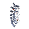 5y4eS S: 精密化の開始モデル |
|---|---|
| 類似構造データ |
-リンク
 PDBj
PDBj


- 集合体
集合体
| 登録構造単位 | 
| ||||||||
|---|---|---|---|---|---|---|---|---|---|
| 1 |
| ||||||||
| 単位格子 |
|
-要素
| #1: タンパク質 | 分子量: 28700.688 Da / 分子数: 1 / 由来タイプ: 組換発現 詳細: The fusion protein of Neurofascin, Linker, and Ankyrin-3 由来: (組換発現) Rattus norvegicus (ドブネズミ) / 遺伝子: Nfasc, Ank3 / 発現宿主:  Escherichia coli (大腸菌) / 参照: UniProt: P97685, UniProt: O70511 Escherichia coli (大腸菌) / 参照: UniProt: P97685, UniProt: O70511 |
|---|---|
| #2: 化合物 | ChemComp-SO4 / 硫酸塩  分子量: 96.063 Da / 分子数: 1 / 由来タイプ: 合成 / 式: SO4 分子量: 96.063 Da / 分子数: 1 / 由来タイプ: 合成 / 式: SO4 |
| #3: 化合物 | ChemComp-GOL / グリセリン  分子量: 92.094 Da / 分子数: 1 / 由来タイプ: 合成 / 式: C3H8O3 分子量: 92.094 Da / 分子数: 1 / 由来タイプ: 合成 / 式: C3H8O3 |
| #4: 水 | ChemComp-HOH / 水 分子量: 18.015 Da / 分子数: 30 / 由来タイプ: 天然 / 式: H2O 分子量: 18.015 Da / 分子数: 30 / 由来タイプ: 天然 / 式: H2O |
| 研究の焦点であるリガンドがあるか | N |
-実験情報
-実験
| 実験 | 手法: X線回折 / 使用した結晶の数: 1 |
|---|
- 試料調製
試料調製
| 結晶 | マシュー密度: 3.72 Å3/Da / 溶媒含有率: 66.91 % |
|---|---|
| 結晶化 | 温度: 289.2 K / 手法: 蒸気拡散法, シッティングドロップ法 / pH: 5.5 詳細: 0.1 M ammonium acetate, 0.1 M Bis-Tris (pH 5.5), 17% w/v polyethylene glycol 10000 |
-データ収集
| 回折 | 平均測定温度: 100 K / Serial crystal experiment: N | |||||||||||||||||||||||||||||||||||||||||||||||||||||||||||||||||||||||||||||||||||||||||||||||||||||||||||||||||||||||||||||||||||||||||||||||||||||||||||||||||||||||||||||||||||||||||||||
|---|---|---|---|---|---|---|---|---|---|---|---|---|---|---|---|---|---|---|---|---|---|---|---|---|---|---|---|---|---|---|---|---|---|---|---|---|---|---|---|---|---|---|---|---|---|---|---|---|---|---|---|---|---|---|---|---|---|---|---|---|---|---|---|---|---|---|---|---|---|---|---|---|---|---|---|---|---|---|---|---|---|---|---|---|---|---|---|---|---|---|---|---|---|---|---|---|---|---|---|---|---|---|---|---|---|---|---|---|---|---|---|---|---|---|---|---|---|---|---|---|---|---|---|---|---|---|---|---|---|---|---|---|---|---|---|---|---|---|---|---|---|---|---|---|---|---|---|---|---|---|---|---|---|---|---|---|---|---|---|---|---|---|---|---|---|---|---|---|---|---|---|---|---|---|---|---|---|---|---|---|---|---|---|---|---|---|---|---|---|---|
| 放射光源 | 由来: シンクロトロン / サイト: SSRF / ビームライン: BL17U1 / 波長: 0.97915 Å | |||||||||||||||||||||||||||||||||||||||||||||||||||||||||||||||||||||||||||||||||||||||||||||||||||||||||||||||||||||||||||||||||||||||||||||||||||||||||||||||||||||||||||||||||||||||||||||
| 検出器 | タイプ: ADSC QUANTUM 315r / 検出器: CCD / 日付: 2017年7月16日 | |||||||||||||||||||||||||||||||||||||||||||||||||||||||||||||||||||||||||||||||||||||||||||||||||||||||||||||||||||||||||||||||||||||||||||||||||||||||||||||||||||||||||||||||||||||||||||||
| 放射 | プロトコル: SINGLE WAVELENGTH / 単色(M)・ラウエ(L): M / 散乱光タイプ: x-ray | |||||||||||||||||||||||||||||||||||||||||||||||||||||||||||||||||||||||||||||||||||||||||||||||||||||||||||||||||||||||||||||||||||||||||||||||||||||||||||||||||||||||||||||||||||||||||||||
| 放射波長 | 波長: 0.97915 Å / 相対比: 1 | |||||||||||||||||||||||||||||||||||||||||||||||||||||||||||||||||||||||||||||||||||||||||||||||||||||||||||||||||||||||||||||||||||||||||||||||||||||||||||||||||||||||||||||||||||||||||||||
| 反射 | 解像度: 2.5→50 Å / Num. obs: 15440 / % possible obs: 99.6 % / 冗長度: 5.4 % / Rmerge(I) obs: 0.124 / Rpim(I) all: 0.059 / Rrim(I) all: 0.137 / Χ2: 2.027 / Net I/σ(I): 8.1 / Num. measured all: 83059 | |||||||||||||||||||||||||||||||||||||||||||||||||||||||||||||||||||||||||||||||||||||||||||||||||||||||||||||||||||||||||||||||||||||||||||||||||||||||||||||||||||||||||||||||||||||||||||||
| 反射 シェル | Diffraction-ID: 1
|
- 解析
解析
| ソフトウェア |
| ||||||||||||||||||||||||||||||||||||||||||
|---|---|---|---|---|---|---|---|---|---|---|---|---|---|---|---|---|---|---|---|---|---|---|---|---|---|---|---|---|---|---|---|---|---|---|---|---|---|---|---|---|---|---|---|
| 精密化 | 構造決定の手法: 分子置換 開始モデル: 5Y4E 解像度: 2.5→34.6 Å / SU ML: 0.3 / 交差検証法: THROUGHOUT / σ(F): 0.32 / 位相誤差: 22.88 / 立体化学のターゲット値: ML
| ||||||||||||||||||||||||||||||||||||||||||
| 溶媒の処理 | 減衰半径: 0.9 Å / VDWプローブ半径: 1.11 Å / 溶媒モデル: FLAT BULK SOLVENT MODEL | ||||||||||||||||||||||||||||||||||||||||||
| 原子変位パラメータ | Biso max: 115.15 Å2 / Biso mean: 50.9493 Å2 / Biso min: 28.42 Å2 | ||||||||||||||||||||||||||||||||||||||||||
| 精密化ステップ | サイクル: final / 解像度: 2.5→34.6 Å
| ||||||||||||||||||||||||||||||||||||||||||
| LS精密化 シェル | Refine-ID: X-RAY DIFFRACTION / Rfactor Rfree error: 0 / Total num. of bins used: 5
|
