 Movie
Movie Controller


+ Open data

- Basic information
| Entry | Database: PDB / ID: 7xc8 | ||||||
|---|---|---|---|---|---|---|---|
| Title | Crystal structure of cotton alpha-like expansin GhEXLA1 | ||||||
 Components Components | Beta-expansin | ||||||
 Keywords Keywords |  PLANT PROTEIN / alpha-like expansin GhEXLA1 / cell wall PLANT PROTEIN / alpha-like expansin GhEXLA1 / cell wall | ||||||
| Function / homology |  Function and homology informationsexual reproduction / anatomical structure morphogenesis / extracellular region Function and homology informationsexual reproduction / anatomical structure morphogenesis / extracellular regionSimilarity search - Function | ||||||
| Biological species |  Gossypium hirsutum (cotton) Gossypium hirsutum (cotton) | ||||||
| Method | X-RAY DIFFRACTION / SYNCHROTRON / MOLECULAR REPLACEMENT / Resolution: 2.55 Å | ||||||
 Authors Authors | Zhao, F. / Men, S. / Xue, Y. / Tu, L.L. / Yin, P. / Zhang, X.L. | ||||||
| Funding support |  China, 1items China, 1items
| ||||||
 Citation Citation | Journal: To Be Published Title: Crystal structure of cotton alpha-like expansin GhEXLA1 Authors: Zhao, F. / Men, S. / Xue, Y. / Tu, L.L. / Yin, P. / Zhang, X.L. | ||||||
| History |
|
- Structure visualization
| Structure viewer | Molecule: MolmilJmol/JSmol |
|---|
- Downloads & links
-Download
| PDBx/mmCIF format | 7xc8.cif.gz | 67.7 KB | Display | PDBx/mmCIF format |
|---|---|---|---|---|
| PDB format | pdb7xc8.ent.gz | 42 KB | Display | PDB format |
| PDBx/mmJSON format | 7xc8.json.gz | Tree view | PDBx/mmJSON format | |
| Others |  Other downloads Other downloads |
-Validation report
| Arichive directory | https://data.pdbj.org/pub/pdb/validation_reports/xc/7xc8ftp://data.pdbj.org/pub/pdb/validation_reports/xc/7xc8 | HTTPS FTP |
|---|
-Related structure data
| Similar structure data |
|---|
-Links
 PDBj
PDBj- Assembly
| Deposited unit | 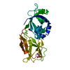
| ||||||||||||
|---|---|---|---|---|---|---|---|---|---|---|---|---|---|
| 1 |
| ||||||||||||
| Unit cell |
| ||||||||||||
| Components on special symmetry positions |
|
-Components
| #1: Protein | Mass: 27048.711 Da / Num. of mol.: 1 Source method: isolated from a genetically manipulated source Source: (gene. exp.) Gossypium hirsutum (cotton) / Gene: LOC107921049, EXPB-L3D / Production host:   Spodoptera frugiperda (fall armyworm) / References: UniProt: A0A1U8L037 Spodoptera frugiperda (fall armyworm) / References: UniProt: A0A1U8L037 | ||||||
|---|---|---|---|---|---|---|---|
| #2: Chemical | Glycerol  Mass: 92.094 Da / Num. of mol.: 2 / Source method: obtained synthetically / Formula: C3H8O3 Mass: 92.094 Da / Num. of mol.: 2 / Source method: obtained synthetically / Formula: C3H8O3#3: Chemical | ChemComp-PEG / | Diethylene glycol  Mass: 106.120 Da / Num. of mol.: 1 / Source method: obtained synthetically / Formula: C4H10O3 Mass: 106.120 Da / Num. of mol.: 1 / Source method: obtained synthetically / Formula: C4H10O3#4: Water | ChemComp-HOH / | Water Mass: 18.015 Da / Num. of mol.: 63 / Source method: isolated from a natural source / Formula: H2O Mass: 18.015 Da / Num. of mol.: 63 / Source method: isolated from a natural source / Formula: H2OHas ligand of interest | N | |
-Experimental details
-Experiment
| Experiment | Method: X-RAY DIFFRACTION / Number of used crystals: 1 |
|---|
- Sample preparation
| Crystal | Density Matthews: 4.06 Å3/Da / Density % sol: 69.74 % |
|---|---|
| Crystal grow | Temperature: 291 K / Method: vapor diffusion, hanging drop / pH: 4 / Details: PEG 3350, Tacsimate pH4.0 / PH range: 3.8-4.5 |
-Data collection
| Diffraction | Mean temperature: 90 K / Serial crystal experiment: N |
|---|---|
| Diffraction source | Source: SYNCHROTRON / Site: SSRF / Beamline: BL19U1 / Wavelength: 0.9785 Å |
| Detector | Type: DECTRIS PILATUS3 6M / Detector: PIXEL / Date: Nov 1, 2021 |
| Radiation | Protocol: SINGLE WAVELENGTH / Monochromatic (M) / Laue (L): M / Scattering type: x-ray |
| Radiation wavelength | Wavelength: 0.9785 Å / Relative weight: 1 |
| Reflection | Resolution: 2.55→19.88 Å / Num. obs: 13869 / % possible obs: 99.8 % / Redundancy: 38 % / Biso Wilson estimate: 44.96 Å2 / CC1/2: 1 / Rmerge(I) obs: 0.124 / Rpim(I) all: 0.028 / Rrim(I) all: 0.127 / Χ2: 0.99 / Net I/σ(I): 33.3 |
| Reflection shell | Resolution: 2.55→2.66 Å / Redundancy: 39 % / Rmerge(I) obs: 0.867 / Mean I/σ(I) obs: 6.3 / Num. unique obs: 1663 / CC1/2: 0.963 / Rpim(I) all: 0.193 / Rrim(I) all: 0.888 / Χ2: 0.99 / % possible all: 100 |
- Processing
| Software |
| |||||||||||||||||||||||||||||||||||||||||||||||||||||||||||||||||||||||||||||
|---|---|---|---|---|---|---|---|---|---|---|---|---|---|---|---|---|---|---|---|---|---|---|---|---|---|---|---|---|---|---|---|---|---|---|---|---|---|---|---|---|---|---|---|---|---|---|---|---|---|---|---|---|---|---|---|---|---|---|---|---|---|---|---|---|---|---|---|---|---|---|---|---|---|---|---|---|---|---|
| Refinement | Method to determine structure: MOLECULAR REPLACEMENT / Resolution: 2.55→19.88 Å / SU ML: 0.3007 / Cross valid method: FREE R-VALUE / σ(F): 1.34 / Phase error: 24.2791 Stereochemistry target values: GeoStd + Monomer Library + CDL v1.2
| |||||||||||||||||||||||||||||||||||||||||||||||||||||||||||||||||||||||||||||
| Solvent computation | Shrinkage radii: 0.9 Å / VDW probe radii: 1.11 Å / Solvent model: FLAT BULK SOLVENT MODEL | |||||||||||||||||||||||||||||||||||||||||||||||||||||||||||||||||||||||||||||
| Displacement parameters | Biso mean: 48.71 Å2 | |||||||||||||||||||||||||||||||||||||||||||||||||||||||||||||||||||||||||||||
| Refinement step | Cycle: LAST / Resolution: 2.55→19.88 Å
| |||||||||||||||||||||||||||||||||||||||||||||||||||||||||||||||||||||||||||||
| Refine LS restraints |
| |||||||||||||||||||||||||||||||||||||||||||||||||||||||||||||||||||||||||||||
| LS refinement shell |
|
