 Movie
Movie Controller


+ Open data

- Basic information
| Entry | Database: PDB / ID: 7x5s | ||||||
|---|---|---|---|---|---|---|---|
| Title | Crystal structure of AtHPPD-Y14157 complex | ||||||
 Components Components | 4-hydroxyphenylpyruvate dioxygenase | ||||||
 Keywords Keywords | OXIDOREDUCTASE / inhibitor / complex | ||||||
| Function / homology |  Function and homology information4-hydroxyphenylpyruvate dioxygenase / 4-hydroxyphenylpyruvate dioxygenase activity / tyrosine catabolic process / L-phenylalanine catabolic process / iron ion binding / identical protein binding / cytoplasm Function and homology information4-hydroxyphenylpyruvate dioxygenase / 4-hydroxyphenylpyruvate dioxygenase activity / tyrosine catabolic process / L-phenylalanine catabolic process / iron ion binding / identical protein binding / cytoplasmSimilarity search - Function | ||||||
| Biological species |  Arabidopsis thaliana (thale cress) Arabidopsis thaliana (thale cress) | ||||||
| Method | X-RAY DIFFRACTION / SYNCHROTRON / MOLECULAR REPLACEMENT / Resolution: 1.706 Å | ||||||
 Authors Authors | Lin, H.-Y. / Dong, J. / Yang, G.-F. | ||||||
| Funding support |  China, 1items China, 1items
| ||||||
 Citation Citation | Journal: Adv Agrochem / Year: 2022 Title: Structural insights of 4-Hydrophenylpyruvate dioxygenase inhibition by structurally diverse small molecules Authors: Dong, J. / Dong, J. / Yu, X.H. / Yan, Y.C. / Nan, J.X. / He, B. / Ye, B.Q. / Yang, W.C. / Lin, H.Y. / Yang, G.F. | ||||||
| History |
|
- Structure visualization
| Structure viewer | Molecule: MolmilJmol/JSmol |
|---|
- Downloads & links
-Download
| PDBx/mmCIF format | 7x5s.cif.gz | 94.7 KB | Display | PDBx/mmCIF format |
|---|---|---|---|---|
| PDB format | pdb7x5s.ent.gz | 67.3 KB | Display | PDB format |
| PDBx/mmJSON format | 7x5s.json.gz | Tree view | PDBx/mmJSON format | |
| Others |  Other downloads Other downloads |
-Validation report
| Arichive directory | https://data.pdbj.org/pub/pdb/validation_reports/x5/7x5sftp://data.pdbj.org/pub/pdb/validation_reports/x5/7x5s | HTTPS FTP |
|---|
-Related structure data
| Related structure data |  7cqrC  7cqsSC  7x5rC  7x5uC  7x5wC  7x5xC 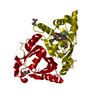 7x6mC  7x6nC  7x7lC 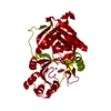 7x8dC  7x8eC 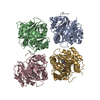 7xntC S: Starting model for refinement C: citing same article ( |
|---|---|
| Similar structure data |
-Links
 PDBj
PDBj
- Assembly
| Deposited unit | 
| ||||||||
|---|---|---|---|---|---|---|---|---|---|
| 1 | 
| ||||||||
| Unit cell |
|
-Components
| #1: Protein | / 4-hydroxyphenylpyruvic acid oxidase / 4HPPD / HPD / HPPDase Mass: 45952.727 Da / Num. of mol.: 1 Source method: isolated from a genetically manipulated source Source: (gene. exp.) Arabidopsis thaliana (thale cress) / Gene: HPD, PDS1, At1g06570, F12K11.9Production host:  Escherichia coli 'BL21-Gold(DE3)pLysS AG' (bacteria) Escherichia coli 'BL21-Gold(DE3)pLysS AG' (bacteria)References: UniProt: P93836, 4-hydroxyphenylpyruvate dioxygenase |
|---|---|
| #2: Chemical | ChemComp-CO /   Mass: 58.933 Da / Num. of mol.: 1 / Source method: obtained synthetically / Formula: Co Mass: 58.933 Da / Num. of mol.: 1 / Source method: obtained synthetically / Formula: Co |
| #3: Chemical | ChemComp-9QL /   Mass: 406.434 Da / Num. of mol.: 1 / Source method: obtained synthetically / Formula: C22H22N4O4 Mass: 406.434 Da / Num. of mol.: 1 / Source method: obtained synthetically / Formula: C22H22N4O4 |
| #4: Water | ChemComp-HOH / Water Mass: 18.015 Da / Num. of mol.: 152 / Source method: isolated from a natural source / Formula: H2O Mass: 18.015 Da / Num. of mol.: 152 / Source method: isolated from a natural source / Formula: H2O |
| Has ligand of interest | N |
-Experimental details
-Experiment
| Experiment | Method: X-RAY DIFFRACTION / Number of used crystals: 1 |
|---|
- Sample preparation
| Crystal | Density Matthews: 2.18 Å3/Da / Density % sol: 43.46 % |
|---|---|
| Crystal grow | Temperature: 293 K / Method: vapor diffusion, hanging drop / pH: 8.5 Details: 0.1M Tris/Bicine pH 8.5, 15% (v/v) MPD, 15% (w/v) PEG 1000, 15% (w/v) PEG 3350, 0.03M NaBr, 0.03M NaF, 0.03M NaI |
-Data collection
| Diffraction | Mean temperature: 100 K / Serial crystal experiment: N |
|---|---|
| Diffraction source | Source: SYNCHROTRON / Site: SSRF / Beamline: BL19U1 / Wavelength: 0.987 Å |
| Detector | Type: ADSC QUANTUM 315r / Detector: CCD / Date: Jun 10, 2021 |
| Radiation | Protocol: SINGLE WAVELENGTH / Monochromatic (M) / Laue (L): M / Scattering type: x-ray |
| Radiation wavelength | Wavelength: 0.987 Å / Relative weight: 1 |
| Reflection | Resolution: 1.7→30 Å / Num. obs: 38655 / % possible obs: 90.5 % / Redundancy: 3.7 % / CC1/2: 0.991 / Rmerge(I) obs: 0.065 / Net I/σ(I): 16.9 |
| Reflection shell | Resolution: 1.7→1.73 Å / Rmerge(I) obs: 0.412 / Mean I/σ(I) obs: 2.4 / Num. unique obs: 1946 / CC1/2: 0.917 |
- Processing
| Software |
| |||||||||||||||||||||||||||||||||||||||||||||||||||||||||||||||||||||||||||||||||||||||||||||||||||||||||
|---|---|---|---|---|---|---|---|---|---|---|---|---|---|---|---|---|---|---|---|---|---|---|---|---|---|---|---|---|---|---|---|---|---|---|---|---|---|---|---|---|---|---|---|---|---|---|---|---|---|---|---|---|---|---|---|---|---|---|---|---|---|---|---|---|---|---|---|---|---|---|---|---|---|---|---|---|---|---|---|---|---|---|---|---|---|---|---|---|---|---|---|---|---|---|---|---|---|---|---|---|---|---|---|---|---|---|
| Refinement | Method to determine structure: MOLECULAR REPLACEMENT Starting model: 7CQS Resolution: 1.706→28.728 Å / SU ML: 0.19 / Cross valid method: FREE R-VALUE / σ(F): 1.38 / Phase error: 28.18 / Stereochemistry target values: ML
| |||||||||||||||||||||||||||||||||||||||||||||||||||||||||||||||||||||||||||||||||||||||||||||||||||||||||
| Solvent computation | Shrinkage radii: 0.9 Å / VDW probe radii: 1.11 Å / Solvent model: FLAT BULK SOLVENT MODEL | |||||||||||||||||||||||||||||||||||||||||||||||||||||||||||||||||||||||||||||||||||||||||||||||||||||||||
| Refinement step | Cycle: LAST / Resolution: 1.706→28.728 Å
| |||||||||||||||||||||||||||||||||||||||||||||||||||||||||||||||||||||||||||||||||||||||||||||||||||||||||
| Refine LS restraints |
| |||||||||||||||||||||||||||||||||||||||||||||||||||||||||||||||||||||||||||||||||||||||||||||||||||||||||
| LS refinement shell |
|
