 Movie
Movie Controller

[English] 日本語
 Yorodumi
- PDB-7x06: CryoEM structure of chitin synthase 1 from Phytophthora sojae com... -
+ Open data

- Basic information
| Entry | Database: PDB / ID: 7x06 | ||||||||||||
|---|---|---|---|---|---|---|---|---|---|---|---|---|---|
| Title | CryoEM structure of chitin synthase 1 from Phytophthora sojae complexed with UDP | ||||||||||||
 Components Components | Chitin synthase | ||||||||||||
 Keywords Keywords | TRANSFERASE / carbohydate / biosynthetic protein / membrane protein | ||||||||||||
| Function / homology |  Function and homology information Function and homology informationchitin biosynthetic process / chitin synthase / chitin synthase activity / membraneSimilarity search - Function | ||||||||||||
| Biological species |  Phytophthora sojae strain P6497 (eukaryote) Phytophthora sojae strain P6497 (eukaryote) | ||||||||||||
| Method | ELECTRON MICROSCOPY / single particle reconstruction / cryo EM / Resolution: 3.1 Å | ||||||||||||
 Authors Authors | Chen, W. / Cao, P. / Gong, Y. / Yang, Q. | ||||||||||||
| Funding support |  China, 3items China, 3items
| ||||||||||||
 Citation Citation | Journal: Nature / Year: 2022 Title: Structural basis for directional chitin biosynthesis. Authors: Wei Chen / Peng Cao / Yuansheng Liu / Ailing Yu / Dong Wang / Lei Chen / Rajamanikandan Sundarraj / Zhiguang Yuchi / Yong Gong / Hans Merzendorfer / Qing Yang /  Abstract: Chitin, the most abundant aminopolysaccharide in nature, is an extracellular polymer consisting of N-acetylglucosamine (GlcNAc) units. The key reactions of chitin biosynthesis are catalysed by chitin ...Chitin, the most abundant aminopolysaccharide in nature, is an extracellular polymer consisting of N-acetylglucosamine (GlcNAc) units. The key reactions of chitin biosynthesis are catalysed by chitin synthase, a membrane-integrated glycosyltransferase that transfers GlcNAc from UDP-GlcNAc to a growing chitin chain. However, the precise mechanism of this process has yet to be elucidated. Here we report five cryo-electron microscopy structures of a chitin synthase from the devastating soybean root rot pathogenic oomycete Phytophthora sojae (PsChs1). They represent the apo, GlcNAc-bound, nascent chitin oligomer-bound, UDP-bound (post-synthesis) and chitin synthase inhibitor nikkomycin Z-bound states of the enzyme, providing detailed views into the multiple steps of chitin biosynthesis and its competitive inhibition. The structures reveal the chitin synthesis reaction chamber that has the substrate-binding site, the catalytic centre and the entrance to the polymer-translocating channel that allows the product polymer to be discharged. This arrangement reflects consecutive key events in chitin biosynthesis from UDP-GlcNAc binding and polymer elongation to the release of the product. We identified a swinging loop within the chitin-translocating channel, which acts as a 'gate lock' that prevents the substrate from leaving while directing the product polymer into the translocating channel for discharge to the extracellular side of the cell membrane. This work reveals the directional multistep mechanism of chitin biosynthesis and provides a structural basis for inhibition of chitin synthesis. | ||||||||||||
| History |
|
- Structure visualization
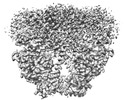
| Structure viewer | Molecule: MolmilJmol/JSmol |
|---|
- Downloads & links
-Download
| PDBx/mmCIF format | 7x06.cif.gz | 287.6 KB | Display | PDBx/mmCIF format |
|---|---|---|---|---|
| PDB format | pdb7x06.ent.gz | 233.9 KB | Display | PDB format |
| PDBx/mmJSON format | 7x06.json.gz | Tree view | PDBx/mmJSON format | |
| Others |  Other downloads Other downloads |
-Validation report
| Arichive directory | https://data.pdbj.org/pub/pdb/validation_reports/x0/7x06ftp://data.pdbj.org/pub/pdb/validation_reports/x0/7x06 | HTTPS FTP |
|---|
-Related structure data
| Related structure data |  32918MC  7wjmC  7wjnC  7wjoC  7x05C C: citing same article ( M: map data used to model this data |
|---|---|
| Similar structure data |


-Links
 PDBj
PDBj


- Assembly
| Deposited unit | 
|
|---|---|
| 1 |
|
-Components
| #1: Protein | Mass: 103146.703 Da / Num. of mol.: 2 Source method: isolated from a genetically manipulated source Source: (gene. exp.) Phytophthora sojae strain P6497 (eukaryote)Strain: P6497 / Gene: PHYSODRAFT_557500 / Cell line (production host): HEK293 / Production host:  Homo sapiens (human) / References: UniProt: G4Z2L3, chitin synthase Homo sapiens (human) / References: UniProt: G4Z2L3, chitin synthase#2: Chemical | Uridine diphosphate  Type: RNA linking / Mass: 404.161 Da / Num. of mol.: 2 / Source method: obtained synthetically / Formula: C9H14N2O12P2 / Comment: UDP*YM Type: RNA linking / Mass: 404.161 Da / Num. of mol.: 2 / Source method: obtained synthetically / Formula: C9H14N2O12P2 / Comment: UDP*YM#3: Chemical |   Mass: 24.305 Da / Num. of mol.: 2 / Source method: obtained synthetically / Formula: Mg Mass: 24.305 Da / Num. of mol.: 2 / Source method: obtained synthetically / Formula: MgHas ligand of interest | N | |
|---|
-Experimental details
-Experiment
| Experiment | Method: ELECTRON MICROSCOPY |
|---|---|
| EM experiment | Aggregation state: PARTICLE / 3D reconstruction method: single particle reconstruction |
- Sample preparation
| Component | Name: Chitin synthase 1 / Type: COMPLEX / Entity ID: #1 / Source: RECOMBINANT |
|---|---|
| Source (natural) | Organism: Phytophthora sojae strain P6497 (eukaryote) |
| Source (recombinant) | Organism: Homo sapiens (human) / Cell: HEK293 |
| Buffer solution | pH: 8 |
| Specimen | Conc.: 5 mg/ml / Embedding applied: NO / Shadowing applied: NO / Staining applied: NO / Vitrification applied: YES |
| Vitrification | Cryogen name: ETHANE |
- Electron microscopy imaging
| Experimental equipment |  Model: Titan Krios / Image courtesy: FEI Company |
|---|---|
| Microscopy | Model: FEI TITAN KRIOS |
| Electron gun | Electron source: FIELD EMISSION GUN / Accelerating voltage: 300 kV / Illumination mode: FLOOD BEAM |
| Electron lens | Mode: BRIGHT FIELDBright-field microscopy / Nominal defocus max: 2000 nm / Nominal defocus min: 1200 nm |
| Image recording | Electron dose: 60 e/Å2 / Film or detector model: GATAN K2 SUMMIT (4k x 4k) |
- Processing
| Software | Name: PHENIX / Version: 1.19.2_4158: / Classification: refinement | ||||||||||||||||||||||||
|---|---|---|---|---|---|---|---|---|---|---|---|---|---|---|---|---|---|---|---|---|---|---|---|---|---|
| CTF correction | Type: PHASE FLIPPING AND AMPLITUDE CORRECTION | ||||||||||||||||||||||||
| Symmetry | Point symmetry: C2 (2 fold cyclic) | ||||||||||||||||||||||||
| 3D reconstruction | Resolution: 3.1 Å / Resolution method: FSC 0.143 CUT-OFF / Num. of particles: 224465 / Symmetry type: POINT | ||||||||||||||||||||||||
| Refine LS restraints |
|
