 Movie
Movie Controller

[English] 日本語
 Yorodumi
- PDB-7wwd: Crystal structure of Saccharomyces cerevisiae Sfh2 complexed with... -
+ Open data

- Basic information
| Entry | Database: PDB / ID: 7wwd | ||||||
|---|---|---|---|---|---|---|---|
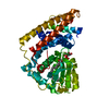
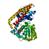
| Title | Crystal structure of Saccharomyces cerevisiae Sfh2 complexed with squalene | ||||||
 Components Components | Phosphatidylinositol transfer protein CSR1 | ||||||
 Keywords Keywords | LIPID TRANSPORT /  Sec14 / phosphatidylinositol / squalene / transport Sec14 / phosphatidylinositol / squalene / transport | ||||||
| Function / homology |  Function and homology information Function and homology informationnegative regulation of phosphatidylglycerol biosynthetic process / positive regulation of phosphatidylcholine biosynthetic process / phosphatidylinositol transfer activity / prospore membrane / phospholipid catabolic process / phosphatidylinositol metabolic process / Golgi to plasma membrane protein transport / negative regulation of fatty acid biosynthetic process / Golgi to plasma membrane transport / phospholipid transport ...negative regulation of phosphatidylglycerol biosynthetic process / positive regulation of phosphatidylcholine biosynthetic process / phosphatidylinositol transfer activity / prospore membrane / phospholipid catabolic process / phosphatidylinositol metabolic process / Golgi to plasma membrane protein transport / negative regulation of fatty acid biosynthetic process / Golgi to plasma membrane transport / phospholipid transport / lipid droplet / endosome / endoplasmic reticulum / mitochondrion / cytosolSimilarity search - Function | ||||||
| Biological species |  Saccharomyces cerevisiae S288C (yeast) Saccharomyces cerevisiae S288C (yeast) | ||||||
| Method | X-RAY DIFFRACTION / SYNCHROTRON / MOLECULAR REPLACEMENT / Resolution: 2.39 Å | ||||||
 Authors Authors | Chen, L. / Tan, L. / Im, Y.J. | ||||||
| Funding support |  Korea, Republic Of, 1items Korea, Republic Of, 1items
| ||||||
 Citation Citation | Journal: Acta Crystallogr D Struct Biol / Year: 2022 Title: Structural basis of ligand recognition and transport by Sfh2, a yeast phosphatidylinositol transfer protein of the Sec14 superfamily. Authors: Chen, L. / Tan, L. / Im, Y.J. | ||||||
| History |
|
- Structure visualization
| Structure viewer | Molecule: MolmilJmol/JSmol |
|---|
- Downloads & links
-Download
| PDBx/mmCIF format | 7wwd.cif.gz | 100.7 KB | Display | PDBx/mmCIF format |
|---|---|---|---|---|
| PDB format | pdb7wwd.ent.gz | 66.7 KB | Display | PDB format |
| PDBx/mmJSON format | 7wwd.json.gz | Tree view | PDBx/mmJSON format | |
| Others |  Other downloads Other downloads |
-Validation report
| Arichive directory | https://data.pdbj.org/pub/pdb/validation_reports/ww/7wwdftp://data.pdbj.org/pub/pdb/validation_reports/ww/7wwd | HTTPS FTP |
|---|
-Related structure data

-Links
 PDBj
PDBj- Assembly
| Deposited unit | 
| ||||||||||||
|---|---|---|---|---|---|---|---|---|---|---|---|---|---|
| 1 |
| ||||||||||||
| Unit cell |
|
-Components
| #1: Protein | Mass: 46570.641 Da / Num. of mol.: 1 / Mutation: deletion of loop residues (44-49 and 61-66) Source method: isolated from a genetically manipulated source Source: (gene. exp.) Saccharomyces cerevisiae S288C (yeast) / Strain: S288c / Gene: CSR1, SFH2, YLR380W / Plasmid: pHIS2-ThrDetails (production host): N-terminal thrombin cleavable hexahistidine-tag Production host:  Escherichia coli BL21(DE3) (bacteria) / References: UniProt: Q06705 Escherichia coli BL21(DE3) (bacteria) / References: UniProt: Q06705 |
|---|---|
| #2: Chemical | ChemComp-SQL / (Squalene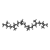  Mass: 410.718 Da / Num. of mol.: 1 / Source method: obtained synthetically / Formula: C30H50 / Feature type: SUBJECT OF INVESTIGATION Mass: 410.718 Da / Num. of mol.: 1 / Source method: obtained synthetically / Formula: C30H50 / Feature type: SUBJECT OF INVESTIGATION |
| #3: Water | ChemComp-HOH / Water Mass: 18.015 Da / Num. of mol.: 88 / Source method: isolated from a natural source / Formula: H2O Mass: 18.015 Da / Num. of mol.: 88 / Source method: isolated from a natural source / Formula: H2O |
| Has ligand of interest | Y |
-Experimental details
-Experiment
| Experiment | Method: X-RAY DIFFRACTION / Number of used crystals: 1 |
|---|
- Sample preparation
| Crystal | Density Matthews: 1.97 Å3/Da / Density % sol: 37.59 % |
|---|---|
| Crystal grow | Temperature: 298 K / Method: vapor diffusion, hanging drop / pH: 7 / Details: 0.1M HEPES pH7.0 0.1M KNO3, 30% PEG 3350 |
-Data collection
| Diffraction | Mean temperature: 110 K / Serial crystal experiment: N |
|---|---|
| Diffraction source | Source: SYNCHROTRON / Site: PAL/PLS / Beamline: 7A (6B, 6C1) / Wavelength: 0.9795 Å |
| Detector | Type: ADSC QUANTUM 270 / Detector: CCD / Date: Jul 26, 2021 |
| Radiation | Protocol: SINGLE WAVELENGTH / Monochromatic (M) / Laue (L): M / Scattering type: x-ray |
| Radiation wavelength | Wavelength: 0.9795 Å / Relative weight: 1 |
| Reflection | Resolution: 2.39→50 Å / Num. obs: 13881 / % possible obs: 97.3 % / Redundancy: 4 % / Biso Wilson estimate: 28.63 Å2 / CC1/2: 0.992 / Rmerge(I) obs: 0.101 / Rpim(I) all: 0.05 / Net I/σ(I): 25.1 |
| Reflection shell | Resolution: 2.39→2.44 Å / Redundancy: 4.1 % / Rmerge(I) obs: 0.416 / Mean I/σ(I) obs: 5.8 / Num. unique obs: 700 / CC1/2: 0.921 / % possible all: 100 |
- Processing
| Software |
| |||||||||||||||||||||||||||||||||||||||||||||||||||||||||||||||||||||||||||||
|---|---|---|---|---|---|---|---|---|---|---|---|---|---|---|---|---|---|---|---|---|---|---|---|---|---|---|---|---|---|---|---|---|---|---|---|---|---|---|---|---|---|---|---|---|---|---|---|---|---|---|---|---|---|---|---|---|---|---|---|---|---|---|---|---|---|---|---|---|---|---|---|---|---|---|---|---|---|---|
| Refinement | Method to determine structure: MOLECULAR REPLACEMENT / Resolution: 2.39→32.66 Å / SU ML: 0.2802 / Cross valid method: FREE R-VALUE / σ(F): 1.34 / Phase error: 27.4807 / Details: AlphaFold Q06705 was used as the starting model.
| |||||||||||||||||||||||||||||||||||||||||||||||||||||||||||||||||||||||||||||
| Solvent computation | Shrinkage radii: 0.9 Å / VDW probe radii: 1.11 Å / Solvent model: FLAT BULK SOLVENT MODEL | |||||||||||||||||||||||||||||||||||||||||||||||||||||||||||||||||||||||||||||
| Displacement parameters | Biso mean: 33.09 Å2 | |||||||||||||||||||||||||||||||||||||||||||||||||||||||||||||||||||||||||||||
| Refinement step | Cycle: LAST / Resolution: 2.39→32.66 Å
| |||||||||||||||||||||||||||||||||||||||||||||||||||||||||||||||||||||||||||||
| Refine LS restraints |
| |||||||||||||||||||||||||||||||||||||||||||||||||||||||||||||||||||||||||||||
| LS refinement shell |
|
