 Movie
Movie Controller

[English] 日本語
 Yorodumi
- PDB-7wt6: Crystal structure of full-length peptidyl-tRNA hydrolase from Myc... -
+ Open data

- Basic information
| Entry | Database: PDB / ID: 7wt6 | |||||||||
|---|---|---|---|---|---|---|---|---|---|---|
| Title | Crystal structure of full-length peptidyl-tRNA hydrolase from Mycobacterium tuberculosis | |||||||||
 Components Components | Peptidyl-tRNA hydrolase Alternative ribosome-rescue factor B Alternative ribosome-rescue factor B | |||||||||
 Keywords Keywords | HYDROLASE / Enzyme classification / Peptidyl-tRNA | |||||||||
| Function / homology |  Function and homology informationpeptidyl-tRNA hydrolase / aminoacyl-tRNA hydrolase activity / translation / plasma membrane / cytoplasm Function and homology informationpeptidyl-tRNA hydrolase / aminoacyl-tRNA hydrolase activity / translation / plasma membrane / cytoplasmSimilarity search - Function | |||||||||
| Biological species |  Mycobacteriaceae bacterium (bacteria) Mycobacteriaceae bacterium (bacteria) | |||||||||
| Method | X-RAY DIFFRACTION / SYNCHROTRON / MOLECULAR REPLACEMENT / Resolution: 1.94 Å | |||||||||
 Authors Authors | Kulandaisamy, R. / Das, U. / Inampudi, K.K. | |||||||||
| Funding support |  India, 2items India, 2items
| |||||||||
 Citation Citation | Journal: To Be Published Title: Crystal structure of full-length peptidyl-tRNA hydrolase from Mycobacterium tuberculosis Authors: Kulandaisamy, R. / Das, U. / Inampudi, K.K. | |||||||||
| History |
|
- Structure visualization
| Structure viewer | Molecule: MolmilJmol/JSmol |
|---|
- Downloads & links
-Download
| PDBx/mmCIF format | 7wt6.cif.gz | 65.3 KB | Display | PDBx/mmCIF format |
|---|---|---|---|---|
| PDB format | pdb7wt6.ent.gz | 36.6 KB | Display | PDB format |
| PDBx/mmJSON format | 7wt6.json.gz | Tree view | PDBx/mmJSON format | |
| Others |  Other downloads Other downloads |
-Validation report
| Arichive directory | https://data.pdbj.org/pub/pdb/validation_reports/wt/7wt6ftp://data.pdbj.org/pub/pdb/validation_reports/wt/7wt6 | HTTPS FTP |
|---|
-Related structure data
| Related structure data | 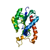 2z2iS S: Starting model for refinement |
|---|---|
| Similar structure data |
-Links
 PDBj
PDBj- Assembly
| Deposited unit | 
| ||||||||||||
|---|---|---|---|---|---|---|---|---|---|---|---|---|---|
| 1 |
| ||||||||||||
| Unit cell |
|
-Components
| #1: Protein | Alternative ribosome-rescue factor B / PTH Mass: 21803.990 Da / Num. of mol.: 1 Source method: isolated from a genetically manipulated source Source: (gene. exp.) Mycobacteriaceae bacterium (bacteria) / Strain: H37Rv / Gene: pth / Production host: Escherichia coli (E. coli) / References: UniProt: P9WHN7, peptidyl-tRNA hydrolase |
|---|---|
| #2: Water | ChemComp-HOH / Water Mass: 18.015 Da / Num. of mol.: 218 / Source method: isolated from a natural source / Formula: H2O Mass: 18.015 Da / Num. of mol.: 218 / Source method: isolated from a natural source / Formula: H2O |
-Experimental details
-Experiment
| Experiment | Method: X-RAY DIFFRACTION / Number of used crystals: 1 |
|---|
- Sample preparation
| Crystal | Density Matthews: 2.15 Å3/Da / Density % sol: 42.85 % |
|---|---|
| Crystal grow | Temperature: 293.15 K / Method: microbatch Details: 25% (W/V) PEG 8000, 0.1M HEPES pH 7.5 5% (V/V) 2-propanol |
-Data collection
| Diffraction | Mean temperature: 100 K / Serial crystal experiment: N |
|---|---|
| Diffraction source | Source: SYNCHROTRON / Site: ESRF  / Beamline: ID30B / Wavelength: 0.976254 Å / Beamline: ID30B / Wavelength: 0.976254 Å |
| Detector | Type: DECTRIS PILATUS3 6M / Detector: PIXEL / Date: Nov 26, 2021 / Details: Vertical CRL/ Horizontal Eliptical mirror |
| Radiation | Monochromator: Si(111) / Protocol: SINGLE WAVELENGTH / Monochromatic (M) / Laue (L): M / Scattering type: x-ray |
| Radiation wavelength | Wavelength: 0.976254 Å / Relative weight: 1 |
| Reflection | Resolution: 1.937→37.201 Å / Num. obs: 13646 / % possible obs: 98.61 % / Redundancy: 8 % / Biso Wilson estimate: 18.92 Å2 / CC1/2: 0.996 / Rmerge(I) obs: 0.149 / Rpim(I) all: 0.055 / Rrim(I) all: 0.159 / Net I/σ(I): 11.4 |
| Reflection shell | Resolution: 1.937→1.97 Å / Redundancy: 6.3 % / Rmerge(I) obs: 0.72 / Mean I/σ(I) obs: 2.5 / Num. unique obs: 587 / CC1/2: 0.887 / Rpim(I) all: 0.29 / Rrim(I) all: 0.78 / % possible all: 87.7 |
- Processing
| Software |
| ||||||||||||||||||||||||||||||||||||||||||
|---|---|---|---|---|---|---|---|---|---|---|---|---|---|---|---|---|---|---|---|---|---|---|---|---|---|---|---|---|---|---|---|---|---|---|---|---|---|---|---|---|---|---|---|
| Refinement | Method to determine structure: MOLECULAR REPLACEMENT Starting model: 2Z2I Resolution: 1.94→37.2 Å / SU ML: 0.2299 / Cross valid method: FREE R-VALUE / σ(F): 1.35 / Phase error: 20.7634 / Stereochemistry target values: CDL v1.2
| ||||||||||||||||||||||||||||||||||||||||||
| Solvent computation | Shrinkage radii: 0.9 Å / VDW probe radii: 1.11 Å / Solvent model: FLAT BULK SOLVENT MODEL | ||||||||||||||||||||||||||||||||||||||||||
| Displacement parameters | Biso mean: 19.58 Å2 | ||||||||||||||||||||||||||||||||||||||||||
| Refinement step | Cycle: LAST / Resolution: 1.94→37.2 Å
| ||||||||||||||||||||||||||||||||||||||||||
| Refine LS restraints |
| ||||||||||||||||||||||||||||||||||||||||||
| LS refinement shell |
|
