 Movie
Movie Controller

[English] 日本語
 Yorodumi
- PDB-7wsj: Crystal structure of the tandem B-box domain of Arabidopsis thali... -
+ Open data

- Basic information
| Entry | Database: PDB / ID: 7wsj | ||||||
|---|---|---|---|---|---|---|---|
| Title | Crystal structure of the tandem B-box domain of Arabidopsis thaliana CONSTANS | ||||||
 Components Components | Zinc finger protein CONSTANS | ||||||
 Keywords Keywords | PLANT PROTEIN / tandem b-box / Zn-coordinating motif / photoperiodic flowering | ||||||
| Function / homology |  Function and homology information Function and homology informationfar-red light signaling pathway / regulation of flower development / response to far red light / flower development / cell differentiation / DNA-binding transcription factor activity / DNA binding / zinc ion binding / nucleusSimilarity search - Function | ||||||
| Biological species |  Arabidopsis thaliana (thale cress) Arabidopsis thaliana (thale cress) | ||||||
| Method | X-RAY DIFFRACTION / SYNCHROTRON / SAD / Resolution: 2.4 Å | ||||||
 Authors Authors | Dahal, P. / Pathak, D. / Kwon, E. / Kim, D.Y. | ||||||
| Funding support |  Korea, Republic Of, 1items Korea, Republic Of, 1items
| ||||||
 Citation Citation | Journal: Biochem.Biophys.Res.Commun. / Year: 2022 Title: Crystal structure of a tandem B-box domain from Arabidopsis CONSTANS. Authors: Dahal, P. / Kwon, E. / Pathak, D. / Kim, D.Y. | ||||||
| History |
|
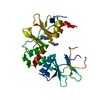
- Structure visualization
| Structure viewer | Molecule: MolmilJmol/JSmol |
|---|
- Downloads & links
-Download
| PDBx/mmCIF format | 7wsj.cif.gz | 122.8 KB | Display | PDBx/mmCIF format |
|---|---|---|---|---|
| PDB format | pdb7wsj.ent.gz | 95.6 KB | Display | PDB format |
| PDBx/mmJSON format | 7wsj.json.gz | Tree view | PDBx/mmJSON format | |
| Others |  Other downloads Other downloads |
-Validation report
| Arichive directory | https://data.pdbj.org/pub/pdb/validation_reports/ws/7wsjftp://data.pdbj.org/pub/pdb/validation_reports/ws/7wsj | HTTPS FTP |
|---|
-Related structure data
| Similar structure data |
|---|


-Links
 PDBj
PDBj
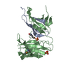
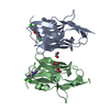
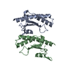
- Assembly
| Deposited unit | 
| ||||||||
|---|---|---|---|---|---|---|---|---|---|
| 1 | 
| ||||||||
| 2 | 
| ||||||||
| Unit cell |
|
-Components
| #1: Protein | Mass: 12012.432 Da / Num. of mol.: 3 Source method: isolated from a genetically manipulated source Source: (gene. exp.) Arabidopsis thaliana (thale cress) / Gene: CO, At5g15840, F14F8_220 / Production host:  Escherichia coli B (bacteria) / References: UniProt: Q39057 Escherichia coli B (bacteria) / References: UniProt: Q39057#2: Chemical | ChemComp-ZN /   Mass: 65.409 Da / Num. of mol.: 12 / Source method: obtained synthetically / Formula: Zn / Feature type: SUBJECT OF INVESTIGATION Mass: 65.409 Da / Num. of mol.: 12 / Source method: obtained synthetically / Formula: Zn / Feature type: SUBJECT OF INVESTIGATION#3: Water | ChemComp-HOH / | Water Mass: 18.015 Da / Num. of mol.: 79 / Source method: isolated from a natural source / Formula: H2O Mass: 18.015 Da / Num. of mol.: 79 / Source method: isolated from a natural source / Formula: H2OHas ligand of interest | Y | |
|---|
-Experimental details
-Experiment
| Experiment | Method: X-RAY DIFFRACTION / Number of used crystals: 1 |
|---|
- Sample preparation
| Crystal | Density Matthews: 3.72 Å3/Da / Density % sol: 66.91 % |
|---|---|
| Crystal grow | Temperature: 293 K / Method: microbatch / pH: 6.5 Details: 1.39 M Lithium Sulfate, 4.55% MPD, 85 mM Imidazole-HCl pH 6.5 |
-Data collection
| Diffraction | Mean temperature: 100 K / Serial crystal experiment: N |
|---|---|
| Diffraction source | Source: SYNCHROTRON / Site: PAL/PLS / Beamline: 11C / Wavelength: 1.28176 Å |
| Detector | Type: DECTRIS PILATUS3 6M / Detector: PIXEL / Date: Jul 8, 2020 |
| Radiation | Protocol: SINGLE WAVELENGTH / Monochromatic (M) / Laue (L): M / Scattering type: x-ray |
| Radiation wavelength | Wavelength: 1.28176 Å / Relative weight: 1 |
| Reflection | Resolution: 2.4→45.93 Å / Num. obs: 19958 / % possible obs: 98.4 % / Redundancy: 3.7 % / Rmerge(I) obs: 0.111 / Net I/σ(I): 7.9 |
| Reflection shell | Resolution: 2.4→2.49 Å / Redundancy: 3.4 % / Rmerge(I) obs: 0.584 / Num. unique obs: 2059 / % possible all: 97.3 |
- Processing
| Software |
| ||||||||||||||||||||||||||||||||||||||||||||||||||||||||
|---|---|---|---|---|---|---|---|---|---|---|---|---|---|---|---|---|---|---|---|---|---|---|---|---|---|---|---|---|---|---|---|---|---|---|---|---|---|---|---|---|---|---|---|---|---|---|---|---|---|---|---|---|---|---|---|---|---|
| Refinement | Method to determine structure: SAD / Resolution: 2.4→45.93 Å / SU ML: 0.29 / Cross valid method: THROUGHOUT / σ(F): 1.96 / Phase error: 25.54 / Stereochemistry target values: ML
| ||||||||||||||||||||||||||||||||||||||||||||||||||||||||
| Solvent computation | Shrinkage radii: 0.9 Å / VDW probe radii: 1.11 Å / Solvent model: FLAT BULK SOLVENT MODEL | ||||||||||||||||||||||||||||||||||||||||||||||||||||||||
| Displacement parameters | Biso max: 147.34 Å2 / Biso mean: 59.3381 Å2 / Biso min: 23.16 Å2 | ||||||||||||||||||||||||||||||||||||||||||||||||||||||||
| Refinement step | Cycle: final / Resolution: 2.4→45.93 Å
| ||||||||||||||||||||||||||||||||||||||||||||||||||||||||
| LS refinement shell | Refine-ID: X-RAY DIFFRACTION / Rfactor Rfree error: 0 / Total num. of bins used: 7
| ||||||||||||||||||||||||||||||||||||||||||||||||||||||||
| Refinement TLS params. | Method: refined / Origin x: 36.2184 Å / Origin y: 23.3896 Å / Origin z: 26.3455 Å
| ||||||||||||||||||||||||||||||||||||||||||||||||||||||||
| Refinement TLS group |
|
