+ Open data
- Basic information
| Entry | Database: PDB / ID: 7wph | ||||||
|---|---|---|---|---|---|---|---|
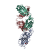
| Title | SARS-CoV2 RBD bound to Fab06 | ||||||
 Components Components |
| ||||||
 Keywords Keywords |  VIRAL PROTEIN/IMMUNE SYSTEM / SARS-Cov2 RBD / VIRAL PROTEIN/IMMUNE SYSTEM / SARS-Cov2 RBD /  antibody / antibody /  PROTEIN BINDING / PROTEIN BINDING /  VIRAL PROTEIN-IMMUNE SYSTEM complex VIRAL PROTEIN-IMMUNE SYSTEM complex | ||||||
| Function / homology |  Function and homology information Function and homology informationMaturation of spike protein / viral translation / Translation of Structural Proteins / Virion Assembly and Release / host cell surface / host extracellular space / suppression by virus of host tetherin activity / Induction of Cell-Cell Fusion / structural constituent of virion / host cell endoplasmic reticulum-Golgi intermediate compartment membrane ...Maturation of spike protein / viral translation / Translation of Structural Proteins / Virion Assembly and Release / host cell surface / host extracellular space / suppression by virus of host tetherin activity / Induction of Cell-Cell Fusion / structural constituent of virion / host cell endoplasmic reticulum-Golgi intermediate compartment membrane / entry receptor-mediated virion attachment to host cell / receptor-mediated endocytosis of virus by host cell / Attachment and Entry /  membrane fusion / positive regulation of viral entry into host cell / receptor-mediated virion attachment to host cell / membrane fusion / positive regulation of viral entry into host cell / receptor-mediated virion attachment to host cell /  receptor ligand activity / host cell surface receptor binding / fusion of virus membrane with host plasma membrane / fusion of virus membrane with host endosome membrane / receptor ligand activity / host cell surface receptor binding / fusion of virus membrane with host plasma membrane / fusion of virus membrane with host endosome membrane /  viral envelope / virion attachment to host cell / SARS-CoV-2 activates/modulates innate and adaptive immune responses / host cell plasma membrane / virion membrane / viral envelope / virion attachment to host cell / SARS-CoV-2 activates/modulates innate and adaptive immune responses / host cell plasma membrane / virion membrane /  membrane / identical protein binding / membrane / identical protein binding /  plasma membrane plasma membraneSimilarity search - Function | ||||||
| Biological species |   Severe acute respiratory syndrome coronavirus 2 Severe acute respiratory syndrome coronavirus 2  Homo sapiens (human) Homo sapiens (human) | ||||||
| Method |  X-RAY DIFFRACTION / X-RAY DIFFRACTION /  SYNCHROTRON / SYNCHROTRON /  MOLECULAR REPLACEMENT / Resolution: 2.89 Å MOLECULAR REPLACEMENT / Resolution: 2.89 Å | ||||||
 Authors Authors | Lin, J.Q. / El Sahili, A. / Lescar, J. | ||||||
| Funding support | 1items
| ||||||
 Citation Citation |  Journal: Nat Commun / Year: 2022 Journal: Nat Commun / Year: 2022Title: Engineering SARS-CoV-2 specific cocktail antibodies into a bispecific format improves neutralizing potency and breadth. Authors: Zhiqiang Ku / Xuping Xie / Jianqing Lin / Peng Gao / Bin Wu / Abbas El Sahili / Hang Su / Yang Liu / Xiaohua Ye / Eddie Yongjun Tan / Xin Li / Xuejun Fan / Boon Chong Goh / Wei Xiong / ...Authors: Zhiqiang Ku / Xuping Xie / Jianqing Lin / Peng Gao / Bin Wu / Abbas El Sahili / Hang Su / Yang Liu / Xiaohua Ye / Eddie Yongjun Tan / Xin Li / Xuejun Fan / Boon Chong Goh / Wei Xiong / Hannah Boyd / Antonio E Muruato / Hui Deng / Hongjie Xia / Jing Zou / Birte K Kalveram / Vineet D Menachery / Ningyan Zhang / Julien Lescar / Pei-Yong Shi / Zhiqiang An /    Abstract: One major limitation of neutralizing antibody-based COVID-19 therapy is the requirement of costly cocktails to reduce emergence of antibody resistance. Here we engineer two bispecific antibodies ...One major limitation of neutralizing antibody-based COVID-19 therapy is the requirement of costly cocktails to reduce emergence of antibody resistance. Here we engineer two bispecific antibodies (bsAbs) using distinct designs and compared them with parental antibodies and their cocktail. Single molecules of both bsAbs block the two epitopes targeted by parental antibodies on the receptor-binding domain (RBD). However, bsAb with the IgG-(scFv) design (14-H-06) but not the CrossMAb design (14-crs-06) shows increased antigen-binding and virus-neutralizing activities against multiple SARS-CoV-2 variants as well as increased breadth of neutralizing activity compared to the cocktail. X-ray crystallography and cryo-EM reveal distinct binding models for individual cocktail antibodies, and computational simulations suggest higher inter-spike crosslinking potentials by 14-H-06 than 14-crs-06. In mouse models of infections by SARS-CoV-2 and multiple variants, 14-H-06 exhibits higher or equivalent therapeutic efficacy than the cocktail. Rationally engineered bsAbs represent a cost-effective alternative to antibody cocktails and a promising strategy to improve potency and breadth. #1: Journal: Biorxiv / Year: 2022 Title: Engineering SARS-CoV-2 cocktail antibodies into a bispecific format improves neutralizing potency and breadth. Authors: Ku, Z. / Xie, X. / Lin, J. / Gao, P. / El Sahili, A. / Su, H. / Liu, Y. / Ye, X. / Li, X. / Fan, X. / Goh, B.C. / Xiong, W. / Boyd, H. / Muruato, A.E. / Deng, H. / Xia, H. / Jing, Z. / ...Authors: Ku, Z. / Xie, X. / Lin, J. / Gao, P. / El Sahili, A. / Su, H. / Liu, Y. / Ye, X. / Li, X. / Fan, X. / Goh, B.C. / Xiong, W. / Boyd, H. / Muruato, A.E. / Deng, H. / Xia, H. / Jing, Z. / Kalveram, B.K. / Menachery, V.D. / Zhang, N. / Lescar, J. / Shi, P.Y. / An, Z. | ||||||
| History |
|
- Structure visualization
| Structure viewer | Molecule:  Molmil Molmil Jmol/JSmol Jmol/JSmol |
|---|
- Downloads & links
- Download
| PDBx/mmCIF format |  7wph.cif.gz 7wph.cif.gz | 272.3 KB | Display |  PDBx/mmCIF format PDBx/mmCIF format |
|---|---|---|---|---|
| PDB format |  pdb7wph.ent.gz pdb7wph.ent.gz | 199.2 KB | Display |  PDB format PDB format |
| PDBx/mmJSON format |  7wph.json.gz 7wph.json.gz | Tree view |  PDBx/mmJSON format PDBx/mmJSON format | |
| Others |  Other downloads Other downloads |
-Validation report
| Arichive directory |  https://data.pdbj.org/pub/pdb/validation_reports/wp/7wph https://data.pdbj.org/pub/pdb/validation_reports/wp/7wph ftp://data.pdbj.org/pub/pdb/validation_reports/wp/7wph ftp://data.pdbj.org/pub/pdb/validation_reports/wp/7wph | HTTPS FTP |
|---|
-Related structure data
| Related structure data |  7wpvC  7xxlC  7c01S S: Starting model for refinement C: citing same article ( |
|---|---|
| Similar structure data | Similarity search - Function & homology  F&H Search F&H Search |
- Links
Links
- Assembly
| Deposited unit | 
| |||||||||||||||||||||||||||||||||||||||||||||||||||||||||||||||||||||||||||||||||||||||||||||||||||||||||||||||
|---|---|---|---|---|---|---|---|---|---|---|---|---|---|---|---|---|---|---|---|---|---|---|---|---|---|---|---|---|---|---|---|---|---|---|---|---|---|---|---|---|---|---|---|---|---|---|---|---|---|---|---|---|---|---|---|---|---|---|---|---|---|---|---|---|---|---|---|---|---|---|---|---|---|---|---|---|---|---|---|---|---|---|---|---|---|---|---|---|---|---|---|---|---|---|---|---|---|---|---|---|---|---|---|---|---|---|---|---|---|---|---|---|
| 1 | 
| |||||||||||||||||||||||||||||||||||||||||||||||||||||||||||||||||||||||||||||||||||||||||||||||||||||||||||||||
| 2 | 
| |||||||||||||||||||||||||||||||||||||||||||||||||||||||||||||||||||||||||||||||||||||||||||||||||||||||||||||||
| Unit cell |
| |||||||||||||||||||||||||||||||||||||||||||||||||||||||||||||||||||||||||||||||||||||||||||||||||||||||||||||||
| Noncrystallographic symmetry (NCS) | NCS domain:
NCS domain segments:
NCS ensembles :
NCS oper:
|
- Components
| #1: Protein | Mass: 30823.617 Da / Num. of mol.: 2 / Fragment: RBD Source method: isolated from a genetically manipulated source Source: (gene. exp.)   Severe acute respiratory syndrome coronavirus 2 Severe acute respiratory syndrome coronavirus 2Gene: S, 2 / Cell line (production host): Expi293 / Production host:   Homo sapiens (human) / References: UniProt: P0DTC2 Homo sapiens (human) / References: UniProt: P0DTC2#2: Antibody | Mass: 22664.959 Da / Num. of mol.: 2 Source method: isolated from a genetically manipulated source Source: (gene. exp.)   Homo sapiens (human) / Cell line (production host): Expi293 / Production host: Homo sapiens (human) / Cell line (production host): Expi293 / Production host:   Homo sapiens (human) Homo sapiens (human)#3: Antibody | Mass: 24114.854 Da / Num. of mol.: 2 Source method: isolated from a genetically manipulated source Source: (gene. exp.)   Homo sapiens (human) / Cell line (production host): Expi293 / Production host: Homo sapiens (human) / Cell line (production host): Expi293 / Production host:   Homo sapiens (human) Homo sapiens (human)#4: Sugar |  N-Acetylglucosamine N-Acetylglucosamine#5: Water | ChemComp-HOH / |  Water WaterHas ligand of interest | N | |
|---|
-Experimental details
-Experiment
| Experiment | Method:  X-RAY DIFFRACTION / Number of used crystals: 1 X-RAY DIFFRACTION / Number of used crystals: 1 |
|---|
- Sample preparation
| Crystal | Density Matthews: 2.42 Å3/Da / Density % sol: 49.25 % / Description: thin plate |
|---|---|
Crystal grow | Temperature: 293.15 K / Method: vapor diffusion, sitting drop / Details: 0.2M magnesium formate dihydrate, 20% w/v PEG 3350 |
-Data collection
| Diffraction | Mean temperature: 80 K / Ambient temp details: liquid nitrogen stream / Serial crystal experiment: N |
|---|---|
| Diffraction source | Source:  SYNCHROTRON / Site: SYNCHROTRON / Site:  Australian Synchrotron Australian Synchrotron  / Beamline: MX2 / Wavelength: 0.9464 Å / Beamline: MX2 / Wavelength: 0.9464 Å |
| Detector | Type: DECTRIS EIGER X 16M / Detector: PIXEL / Date: Jun 30, 2021 |
| Radiation | Protocol: SINGLE WAVELENGTH / Monochromatic (M) / Laue (L): M / Scattering type: x-ray |
| Radiation wavelength | Wavelength : 0.9464 Å / Relative weight: 1 : 0.9464 Å / Relative weight: 1 |
| Reflection | Resolution: 2.89→55 Å / Num. obs: 34904 / % possible obs: 99.81 % / Redundancy: 10.4 % / Biso Wilson estimate: 63.21 Å2 / CC1/2: 0.994 / CC star: 0.998 / Rpim(I) all: 0.1006 / Rrim(I) all: 0.3316 / Net I/σ(I): 8.31 |
| Reflection shell | Resolution: 2.89→2.993 Å / Mean I/σ(I) obs: 1.22 / Num. unique obs: 3389 / CC1/2: 0.522 / % possible all: 96.7 |
- Processing
| Software |
| ||||||||||||||||||||||||||||||||||||||||||||||||||||||||||||||||||||||||||||||||||||||||||||||||||||||||||||||||||||||||||||||||||||||||||||||||||||||||||||||||||||||||
|---|---|---|---|---|---|---|---|---|---|---|---|---|---|---|---|---|---|---|---|---|---|---|---|---|---|---|---|---|---|---|---|---|---|---|---|---|---|---|---|---|---|---|---|---|---|---|---|---|---|---|---|---|---|---|---|---|---|---|---|---|---|---|---|---|---|---|---|---|---|---|---|---|---|---|---|---|---|---|---|---|---|---|---|---|---|---|---|---|---|---|---|---|---|---|---|---|---|---|---|---|---|---|---|---|---|---|---|---|---|---|---|---|---|---|---|---|---|---|---|---|---|---|---|---|---|---|---|---|---|---|---|---|---|---|---|---|---|---|---|---|---|---|---|---|---|---|---|---|---|---|---|---|---|---|---|---|---|---|---|---|---|---|---|---|---|---|---|---|---|
| Refinement | Method to determine structure : :  MOLECULAR REPLACEMENT MOLECULAR REPLACEMENTStarting model: 7C01 Resolution: 2.89→48.15 Å / SU ML: 0.5441 / Cross valid method: FREE R-VALUE / σ(F): 1.25 / Phase error: 31.178 Stereochemistry target values: GeoStd + Monomer Library + CDL v1.2
| ||||||||||||||||||||||||||||||||||||||||||||||||||||||||||||||||||||||||||||||||||||||||||||||||||||||||||||||||||||||||||||||||||||||||||||||||||||||||||||||||||||||||
| Solvent computation | Shrinkage radii: 0.9 Å / VDW probe radii: 1.11 Å / Solvent model: FLAT BULK SOLVENT MODEL | ||||||||||||||||||||||||||||||||||||||||||||||||||||||||||||||||||||||||||||||||||||||||||||||||||||||||||||||||||||||||||||||||||||||||||||||||||||||||||||||||||||||||
| Displacement parameters | Biso mean: 67.21 Å2 | ||||||||||||||||||||||||||||||||||||||||||||||||||||||||||||||||||||||||||||||||||||||||||||||||||||||||||||||||||||||||||||||||||||||||||||||||||||||||||||||||||||||||
| Refinement step | Cycle: LAST / Resolution: 2.89→48.15 Å
| ||||||||||||||||||||||||||||||||||||||||||||||||||||||||||||||||||||||||||||||||||||||||||||||||||||||||||||||||||||||||||||||||||||||||||||||||||||||||||||||||||||||||
| Refine LS restraints |
| ||||||||||||||||||||||||||||||||||||||||||||||||||||||||||||||||||||||||||||||||||||||||||||||||||||||||||||||||||||||||||||||||||||||||||||||||||||||||||||||||||||||||
| Refine LS restraints NCS |
| ||||||||||||||||||||||||||||||||||||||||||||||||||||||||||||||||||||||||||||||||||||||||||||||||||||||||||||||||||||||||||||||||||||||||||||||||||||||||||||||||||||||||
| LS refinement shell |
|
 Movie
Movie Controller


 PDBj
PDBj


