 Movie
Movie Controller


+ Open data

- Basic information
| Entry | Database: PDB / ID: 7wmp | |||||||||
|---|---|---|---|---|---|---|---|---|---|---|
| Title | Tail structure of Helicobacter pylori bacteriophage KHP30 | |||||||||
 Components Components |
| |||||||||
 Keywords Keywords |  VIRAL PROTEIN / PHAGE / PHAGE TAIL / CRYOEM / VIRUS VIRAL PROTEIN / PHAGE / PHAGE TAIL / CRYOEM / VIRUS | |||||||||
| Function / homology | symbiont genome ejection through host cell envelope, short tail mechanism / viral capsid / Orphan protein / Phage tail assembly protein / Portal protein Function and homology information Function and homology information | |||||||||
| Biological species |  Helicobacter phage KHP30 (virus) Helicobacter phage KHP30 (virus) | |||||||||
| Method | ELECTRON MICROSCOPY / single particle reconstruction / cryo EM / Resolution: 3.6 Å | |||||||||
 Authors Authors | Kamiya, R. / Uchiyama, J. / Matsuzaki, S. / Murata, K. / Iwasaki, K. / Miyazaki, N. | |||||||||
| Funding support |  Japan, 2items Japan, 2items
| |||||||||
 Citation Citation | Journal: To Be Published Title: Cryo-EM structure of Helicobacter pylori bacteriophage KHP30 Authors: Kamiya, R. / Uchiyama, J. / Matsuzaki, S. / Murata, K. / Iwasaki, K. / Miyazaki, N. | |||||||||
| History |
|
- Structure visualization
| Structure viewer | Molecule: MolmilJmol/JSmol |
|---|
- Downloads & links
-Download
| PDBx/mmCIF format | 7wmp.cif.gz | 2 MB | Display | PDBx/mmCIF format |
|---|---|---|---|---|
| PDB format | pdb7wmp.ent.gz | 1.7 MB | Display | PDB format |
| PDBx/mmJSON format | 7wmp.json.gz | Tree view | PDBx/mmJSON format | |
| Others |  Other downloads Other downloads |
-Validation report
| Arichive directory | https://data.pdbj.org/pub/pdb/validation_reports/wm/7wmpftp://data.pdbj.org/pub/pdb/validation_reports/wm/7wmp | HTTPS FTP |
|---|
-Related structure data
| Related structure data | 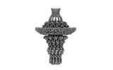 32616MC M: map data used to model this data C: citing same article ( |
|---|---|
| Similar structure data |
-Links
 PDBj
PDBj- Assembly
| Deposited unit | 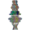
|
|---|---|
| 1 |
|
-Components
| #1: Protein | Mass: 69634.734 Da / Num. of mol.: 12 / Source method: isolated from a natural source / Source: (natural) Helicobacter phage KHP30 (virus) / References: UniProt: I7HHN4#2: Protein | AdapterMass: 22609.309 Da / Num. of mol.: 12 / Source method: isolated from a natural source / Source: (natural) Helicobacter phage KHP30 (virus) / References: UniProt: I7HHN3#3: Protein | Mass: 29741.775 Da / Num. of mol.: 12 / Source method: isolated from a natural source / Source: (natural) Helicobacter phage KHP30 (virus) / References: UniProt: I7HFX1 |
|---|
-Experimental details
-Experiment
| Experiment | Method: ELECTRON MICROSCOPY |
|---|---|
| EM experiment | Aggregation state: PARTICLE / 3D reconstruction method: single particle reconstruction |
- Sample preparation
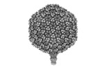
| Component | Name: Helicobacter phage KHP / Type: VIRUS / Entity ID: all / Source: NATURAL |
|---|---|
| Molecular weight | Experimental value: NO |
| Source (natural) | Organism: Helicobacter phage KHP (virus) |
| Details of virus | Empty: NO / Enveloped: NO / Isolate: OTHER / Type: VIRION |
| Virus shell | Name: Head / Diameter: 700 nm / Triangulation number (T number): 9 |
| Buffer solution | pH: 7.2 |
| Specimen | Embedding applied: NO / Shadowing applied: NO / Staining applied: NO / Vitrification applied: YES |
| Vitrification | Instrument: FEI VITROBOT MARK IV / Cryogen name: ETHANE / Humidity: 100 % / Chamber temperature: 277 K |
- Electron microscopy imaging
| Experimental equipment |  Model: Titan Krios / Image courtesy: FEI Company |
|---|---|
| Microscopy | Model: FEI TITAN KRIOS |
| Electron gun | Electron source: FIELD EMISSION GUN / Accelerating voltage: 300 kV / Illumination mode: FLOOD BEAM |
| Electron lens | Mode: BRIGHT FIELDBright-field microscopy / Nominal defocus max: 2500 nm / Nominal defocus min: 1000 nm / Cs: 2.7 mm |
| Specimen holder | Cryogen: NITROGEN / Specimen holder model: FEI TITAN KRIOS AUTOGRID HOLDER |
| Image recording | Average exposure time: 2.5 sec. / Electron dose: 50 e/Å2 / Detector mode: INTEGRATING / Film or detector model: FEI FALCON III (4k x 4k) |
- Processing
| EM software | Name: RELION / Version: 3 / Category: 3D reconstruction |
|---|---|
| CTF correction | Type: PHASE FLIPPING AND AMPLITUDE CORRECTION |
| Particle selection | Num. of particles selected: 32655 |
| Symmetry | Point symmetry: C12 (12 fold cyclic) |
| 3D reconstruction | Resolution: 3.6 Å / Resolution method: FSC 0.143 CUT-OFF / Num. of particles: 27422 / Symmetry type: POINT |
