Entry Database : PDB / ID : 7whhTitle Crystal structure of SARS-CoV-2 omicron RBD and human ACE2 Processed angiotensin-converting enzyme 2 Spike glycoprotein Keywords / / / / / / / Function / homology Function Domain/homology Component
/ / / / / / / / / / / / / / / / / / / / / / / / / / / / / / / / / / / / / / / / / / / / / / / / / / / / / / / / / / / / / / / / / / / / / / / / / / / / / / / / / / / / / / / / / / / / / / / / / / / / / / / / / / / / / / / / / / / Biological species Homo sapiens (human)Method / / / Resolution : 2.6 Å Authors Wang, X.Q. / Lan, J. Funding support Organization Grant number Country National Natural Science Foundation of China (NSFC)
Journal : To Be Published Title : Crystal structure of SARS-CoV-2 omicron RBD and human ACE2Authors : Wang, X.Q. / Lan, J. / Ge, J.W. History Deposition Dec 30, 2021 Deposition site / Processing site Revision 1.0 Jun 1, 2022 Provider / Type Revision 1.1 Nov 29, 2023 Group / Refinement descriptionCategory / chem_comp_bond / pdbx_initial_refinement_model
Show all Show less
 Movie
Movie Controller


 Open data
 Basic information
 Components
 Keywords
 SARS-CoV-2 / spike /
  Function and homology information


 Authors
 China, 1items
  Citation
 Structure visualization
 Downloads & links
 Other downloads
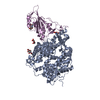
 PDBj
PDBj


 Assembly


 Type: D-saccharide, beta linking / Mass: 221.208 Da / Num. of mol.: 4 / Source method: obtained synthetically / Formula: C8H15NO6


 Mass: 65.409 Da / Num. of mol.: 1 / Source method: obtained synthetically / Formula: Zn
 Mass: 35.453 Da / Num. of mol.: 1 / Source method: obtained synthetically / Formula: Cl
 Sample preparation
 Processing