 Movie
Movie Controller


+ Open data

- Basic information
| Entry | Database: PDB / ID: 7wdw | ||||||
|---|---|---|---|---|---|---|---|
| Title | DsyB in complex with SAH and MTHB | ||||||
 Components Components | DSYB | ||||||
 Keywords Keywords |  TRANSFERASE / Complex / SAM-dependent methyltransferase TRANSFERASE / Complex / SAM-dependent methyltransferase | ||||||
| Function / homology | 4-methylsulfanyl-2-oxidanyl-butanoic acid / S-ADENOSYL-L-HOMOCYSTEINE Function and homology information Function and homology information | ||||||
| Biological species |  Nisaea denitrificans DSM 18348 (bacteria) Nisaea denitrificans DSM 18348 (bacteria) | ||||||
| Method | X-RAY DIFFRACTION / SYNCHROTRON / MOLECULAR REPLACEMENT / Resolution: 2.39 Å | ||||||
 Authors Authors | Li, C.Y. | ||||||
| Funding support | 1items
| ||||||
 Citation Citation | Journal: Mlife / Year: 2022 Title: Mechanistic insights into the key marine dimethylsulfoniopropionate synthesis enzyme DsyB/DSYB. Authors: Li, C.Y. / Crack, J.C. / Newton-Payne, S. / Murphy, A.R.J. / Chen, X.L. / Pinchbeck, B.J. / Zhou, S. / Williams, B.T. / Peng, M. / Zhang, X.H. / Chen, Y. | ||||||
| History |
|
- Structure visualization
| Structure viewer | Molecule: MolmilJmol/JSmol |
|---|
- Downloads & links
-Download
| PDBx/mmCIF format | 7wdw.cif.gz | 267.8 KB | Display | PDBx/mmCIF format |
|---|---|---|---|---|
| PDB format | pdb7wdw.ent.gz | 215 KB | Display | PDB format |
| PDBx/mmJSON format | 7wdw.json.gz | Tree view | PDBx/mmJSON format | |
| Others |  Other downloads Other downloads |
-Validation report
| Arichive directory | https://data.pdbj.org/pub/pdb/validation_reports/wd/7wdwftp://data.pdbj.org/pub/pdb/validation_reports/wd/7wdw | HTTPS FTP |
|---|
-Related structure data
| Related structure data |  7wdqC  7dfd S: Starting model for refinement C: citing same article ( |
|---|---|
| Similar structure data |
-Links
 PDBj
PDBj- Assembly
| Deposited unit | 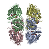
| ||||||||
|---|---|---|---|---|---|---|---|---|---|
| 1 | 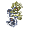
| ||||||||
| 2 | 
| ||||||||
| Unit cell |
|
-Components
| #1: Protein | Mass: 37014.828 Da / Num. of mol.: 4 Source method: isolated from a genetically manipulated source Source: (gene. exp.) Nisaea denitrificans DSM 18348 (bacteria)Production host: Escherichia coli BL21(DE3) (bacteria)#2: Chemical | ChemComp-SAH / S-Adenosyl-L-homocysteine  Type: L-peptide linking / Mass: 384.411 Da / Num. of mol.: 4 / Source method: obtained synthetically / Formula: C14H20N6O5S / Feature type: SUBJECT OF INVESTIGATION Type: L-peptide linking / Mass: 384.411 Da / Num. of mol.: 4 / Source method: obtained synthetically / Formula: C14H20N6O5S / Feature type: SUBJECT OF INVESTIGATION#3: Chemical | ChemComp-H9L / 2-Hydroxy-4-(methylthio)butyric acid  Mass: 150.196 Da / Num. of mol.: 4 / Source method: obtained synthetically / Formula: C5H10O3S / Feature type: SUBJECT OF INVESTIGATION Mass: 150.196 Da / Num. of mol.: 4 / Source method: obtained synthetically / Formula: C5H10O3S / Feature type: SUBJECT OF INVESTIGATION#4: Chemical | ChemComp-TRS / Tris  Mass: 122.143 Da / Num. of mol.: 4 / Source method: obtained synthetically / Formula: C4H12NO3 / Feature type: SUBJECT OF INVESTIGATION / Comment: pH buffer*YM Mass: 122.143 Da / Num. of mol.: 4 / Source method: obtained synthetically / Formula: C4H12NO3 / Feature type: SUBJECT OF INVESTIGATION / Comment: pH buffer*YM#5: Water | ChemComp-HOH / | Water Mass: 18.015 Da / Num. of mol.: 286 / Source method: isolated from a natural source / Formula: H2O Mass: 18.015 Da / Num. of mol.: 286 / Source method: isolated from a natural source / Formula: H2OHas ligand of interest | Y | |
|---|
-Experimental details
-Experiment
| Experiment | Method: X-RAY DIFFRACTION / Number of used crystals: 1 |
|---|
- Sample preparation
| Crystal | Density Matthews: 2.15 Å3/Da / Density % sol: 42.78 % |
|---|---|
| Crystal grow | Temperature: 293 K / Method: vapor diffusion, hanging drop / Details: 0.2 M NaCl, 0.1 M Tris (pH 8.0) and 20% PEG 4000 |
-Data collection
| Diffraction | Mean temperature: 100 K / Serial crystal experiment: N |
|---|---|
| Diffraction source | Source: SYNCHROTRON / Site: SSRF  / Beamline: BL18U1 / Wavelength: 0.979 Å / Beamline: BL18U1 / Wavelength: 0.979 Å |
| Detector | Type: DECTRIS PILATUS 6M / Detector: PIXEL / Date: Dec 5, 2015 |
| Radiation | Protocol: SINGLE WAVELENGTH / Monochromatic (M) / Laue (L): M / Scattering type: x-ray |
| Radiation wavelength | Wavelength: 0.979 Å / Relative weight: 1 |
| Reflection | Resolution: 2.39→50 Å / Num. obs: 49532 / % possible obs: 99.7 % / Redundancy: 3.7 % / Biso Wilson estimate: 32.77 Å2 / Rmerge(I) obs: 0.094 / Χ2: 2.542 / Net I/σ(I): 11.6 |
| Reflection shell | Resolution: 2.4→2.49 Å / Redundancy: 3.7 % / Rmerge(I) obs: 0.457 / Num. unique obs: 5100 / Χ2: 3.175 / % possible all: 99.7 |
- Processing
| Software |
| ||||||||||||||||||||||||||||||||||||||||||||||||||||||||||||||||||||||||||||||||||||||||||||||||||||||||||||||||||||||||||||||
|---|---|---|---|---|---|---|---|---|---|---|---|---|---|---|---|---|---|---|---|---|---|---|---|---|---|---|---|---|---|---|---|---|---|---|---|---|---|---|---|---|---|---|---|---|---|---|---|---|---|---|---|---|---|---|---|---|---|---|---|---|---|---|---|---|---|---|---|---|---|---|---|---|---|---|---|---|---|---|---|---|---|---|---|---|---|---|---|---|---|---|---|---|---|---|---|---|---|---|---|---|---|---|---|---|---|---|---|---|---|---|---|---|---|---|---|---|---|---|---|---|---|---|---|---|---|---|---|
| Refinement | Method to determine structure: MOLECULAR REPLACEMENT Starting model: 7DFD 7dfd Resolution: 2.39→37.16 Å / SU ML: 0.34 / Cross valid method: THROUGHOUT / σ(F): 0 / Phase error: 30 / Stereochemistry target values: ML
| ||||||||||||||||||||||||||||||||||||||||||||||||||||||||||||||||||||||||||||||||||||||||||||||||||||||||||||||||||||||||||||||
| Solvent computation | Shrinkage radii: 0.95 Å / VDW probe radii: 1.2 Å / Solvent model: FLAT BULK SOLVENT MODEL / Bsol: 23.44 Å2 / ksol: 0.3 e/Å3 | ||||||||||||||||||||||||||||||||||||||||||||||||||||||||||||||||||||||||||||||||||||||||||||||||||||||||||||||||||||||||||||||
| Displacement parameters | Biso max: 97.55 Å2 / Biso mean: 43.02 Å2 / Biso min: 19.33 Å2
| ||||||||||||||||||||||||||||||||||||||||||||||||||||||||||||||||||||||||||||||||||||||||||||||||||||||||||||||||||||||||||||||
| Refinement step | Cycle: final / Resolution: 2.39→37.16 Å
| ||||||||||||||||||||||||||||||||||||||||||||||||||||||||||||||||||||||||||||||||||||||||||||||||||||||||||||||||||||||||||||||
| LS refinement shell | Refine-ID: X-RAY DIFFRACTION / Rfactor Rfree error: 0 / Total num. of bins used: 17
|
