 Movie
Movie Controller

[English] 日本語
 Yorodumi
- PDB-7wcy: Crystal Structure of H-2Kb with Cryptosporidium parvum gp40/15 epitope -
+ Open data

- Basic information
| Entry | Database: PDB / ID: 7wcy | ||||||
|---|---|---|---|---|---|---|---|
| Title | Crystal Structure of H-2Kb with Cryptosporidium parvum gp40/15 epitope | ||||||
 Components Components |
| ||||||
 Keywords Keywords |  IMMUNE SYSTEM / MHC I / peptide epitope / T cell immunity IMMUNE SYSTEM / MHC I / peptide epitope / T cell immunity | ||||||
| Function / homology |  Function and homology information Function and homology informationEndosomal/Vacuolar pathway / DAP12 interactions / Antigen Presentation: Folding, assembly and peptide loading of class I MHC / ER-Phagosome pathway / DAP12 signaling / Immunoregulatory interactions between a Lymphoid and a non-Lymphoid cell / regulation of membrane depolarization / antigen processing and presentation of exogenous peptide antigen via MHC class I / inner ear development / antigen processing and presentation of endogenous peptide antigen via MHC class I via ER pathway, TAP-dependent ...Endosomal/Vacuolar pathway / DAP12 interactions / Antigen Presentation: Folding, assembly and peptide loading of class I MHC / ER-Phagosome pathway / DAP12 signaling / Immunoregulatory interactions between a Lymphoid and a non-Lymphoid cell / regulation of membrane depolarization / antigen processing and presentation of exogenous peptide antigen via MHC class I / inner ear development / antigen processing and presentation of endogenous peptide antigen via MHC class I via ER pathway, TAP-dependent / cellular defense response / beta-2-microglobulin binding / Neutrophil degranulation / antigen processing and presentation of endogenous peptide antigen via MHC class I via ER pathway, TAP-independent / antigen processing and presentation of endogenous peptide antigen via MHC class Ib / lumenal side of endoplasmic reticulum membrane / peptide binding / cellular response to iron(III) ion / antigen processing and presentation of exogenous protein antigen via MHC class Ib, TAP-dependent / negative regulation of forebrain neuron differentiation / regulation of erythrocyte differentiation / peptide antigen assembly with MHC class I protein complex / response to molecule of bacterial origin / regulation of iron ion transport / MHC class I peptide loading complex / HFE-transferrin receptor complex / T cell mediated cytotoxicity / cellular response to iron ion / antigen processing and presentation of endogenous peptide antigen via MHC class I / positive regulation of T cell cytokine production / MHC class I protein complex / multicellular organismal-level iron ion homeostasis / negative regulation of neurogenesis / peptide antigen assembly with MHC class II protein complex / positive regulation of receptor-mediated endocytosis / MHC class II protein complex / cellular response to nicotine / positive regulation of T cell mediated cytotoxicity / phagocytic vesicle membrane / peptide antigen binding / positive regulation of cellular senescence / antigen processing and presentation of exogenous peptide antigen via MHC class II / negative regulation of epithelial cell proliferation / positive regulation of immune response / antimicrobial humoral immune response mediated by antimicrobial peptide / sensory perception of smell / positive regulation of T cell activation / negative regulation of neuron projection development / MHC class II protein complex binding / late endosome membrane / T cell differentiation in thymus / iron ion transport / protein refolding / antibacterial humoral response / protein homotetramerization / intracellular iron ion homeostasis / cellular response to lipopolysaccharide / defense response to Gram-negative bacterium / amyloid fibril formation / learning or memory / defense response to Gram-positive bacterium / defense response to bacterium / immune response / lysosomal membrane / external side of plasma membrane / signaling receptor binding / innate immune response / protein-containing complex binding / structural molecule activity / Golgi apparatus / protein homodimerization activity / extracellular space / identical protein binding / plasma membrane / cytosolSimilarity search - Function | ||||||
| Biological species |  Mus musculus (house mouse) Mus musculus (house mouse)  Cryptosporidium parvum (eukaryote) Cryptosporidium parvum (eukaryote) | ||||||
| Method | X-RAY DIFFRACTION / MOLECULAR REPLACEMENT / Resolution: 2.36 Å | ||||||
 Authors Authors | Wang, Y.L. / Gao, M.H. / Zhang, L.X. / Fan, S.H. | ||||||
| Funding support |  China, 1items China, 1items
| ||||||
 Citation Citation | Journal: Mbio / Year: 2023 Title: Structural Analyses of a Dominant Cryptosporidium parvum Epitope Presented by H-2K b Offer New Options To Combat Cryptosporidiosis. Authors: Wang, Y. / Gao, M. / Li, X. / Zhu, W. / Zhao, M. / Li, J. / Liu, X. / Cao, L. / Li, S. / Zhang, S. / Zhang, L. / Fan, S. | ||||||
| History |
|
- Structure visualization
| Structure viewer | Molecule: MolmilJmol/JSmol |
|---|
- Downloads & links
-Download
| PDBx/mmCIF format | 7wcy.cif.gz | 170.3 KB | Display | PDBx/mmCIF format |
|---|---|---|---|---|
| PDB format | pdb7wcy.ent.gz | 133.2 KB | Display | PDB format |
| PDBx/mmJSON format | 7wcy.json.gz | Tree view | PDBx/mmJSON format | |
| Others |  Other downloads Other downloads |
-Validation report
| Arichive directory | https://data.pdbj.org/pub/pdb/validation_reports/wc/7wcyftp://data.pdbj.org/pub/pdb/validation_reports/wc/7wcy | HTTPS FTP |
|---|
-Related structure data
| Related structure data | 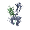 1vacS S: Starting model for refinement |
|---|---|
| Similar structure data |
-Links
 PDBj
PDBj


- Assembly
| Deposited unit | 
| ||||||||
|---|---|---|---|---|---|---|---|---|---|
| 1 | 
| ||||||||
| 2 | 
| ||||||||
| Unit cell |
|
-Components
| #1: Protein | Mass: 31777.438 Da / Num. of mol.: 2 Source method: isolated from a genetically manipulated source Source: (gene. exp.) Mus musculus (house mouse) / Gene: H2-K1, H2-K / Production host:  Escherichia coli (E. coli) / References: UniProt: P01901 Escherichia coli (E. coli) / References: UniProt: P01901#2: Protein | Beta-2 microglobulinMass: 11660.350 Da / Num. of mol.: 2 Source method: isolated from a genetically manipulated source Source: (gene. exp.) Mus musculus (house mouse) / Gene: B2m / Production host: Escherichia coli (E. coli) / References: UniProt: P01887#3: Protein/peptide | Mass: 938.120 Da / Num. of mol.: 2 / Source method: obtained synthetically / Source: (synth.) Cryptosporidium parvum (eukaryote)#4: Chemical | ChemComp-NI / Nickel  Mass: 58.693 Da / Num. of mol.: 8 / Source method: obtained synthetically / Formula: Ni Mass: 58.693 Da / Num. of mol.: 8 / Source method: obtained synthetically / Formula: Ni#5: Water | ChemComp-HOH / | Water Mass: 18.015 Da / Num. of mol.: 60 / Source method: isolated from a natural source / Formula: H2O Mass: 18.015 Da / Num. of mol.: 60 / Source method: isolated from a natural source / Formula: H2OHas ligand of interest | N | |
|---|
-Experimental details
-Experiment
| Experiment | Method: X-RAY DIFFRACTION / Number of used crystals: 1 |
|---|
- Sample preparation
| Crystal | Density Matthews: 2.77 Å3/Da / Density % sol: 55.61 % |
|---|---|
| Crystal grow | Temperature: 291 K / Method: vapor diffusion, sitting drop / pH: 7.5 Details: 12%(w/v) Polyethylene glycol 3350, 0.005M Cobalt(II) chloride hexahydrate; 0.005M Nickel(II) chloride hexahydrate; 0.005M Cadmium chloride hydrate; 0.1M HEPES (PH7.5) |
-Data collection
| Diffraction | Mean temperature: 100 K / Serial crystal experiment: N |
|---|---|
| Diffraction source | Source: ROTATING ANODE / Type: RIGAKU MICROMAX-007 HF / Wavelength: 0.97915 Å |
| Detector | Type: MAR scanner 300 mm plate / Detector: IMAGE PLATE / Date: Nov 20, 2021 |
| Radiation | Protocol: SINGLE WAVELENGTH / Monochromatic (M) / Laue (L): M / Scattering type: x-ray |
| Radiation wavelength | Wavelength: 0.97915 Å / Relative weight: 1 |
| Reflection | Resolution: 2.36→49.47 Å / Num. obs: 40181 / % possible obs: 97.98 % / Redundancy: 2.5 % / CC1/2: 0.999 / Net I/σ(I): 15.259 |
| Reflection shell | Resolution: 2.36→2.45 Å / Num. unique obs: 4091 / CC1/2: 0.826 |
- Processing
| Software |
| ||||||||||||||||||||||||||||||||||||||||||||||||||||||||||||||||||||||||||||||||||||||||||||||||||||||||||||||||||||||||||||||||||||||||||||
|---|---|---|---|---|---|---|---|---|---|---|---|---|---|---|---|---|---|---|---|---|---|---|---|---|---|---|---|---|---|---|---|---|---|---|---|---|---|---|---|---|---|---|---|---|---|---|---|---|---|---|---|---|---|---|---|---|---|---|---|---|---|---|---|---|---|---|---|---|---|---|---|---|---|---|---|---|---|---|---|---|---|---|---|---|---|---|---|---|---|---|---|---|---|---|---|---|---|---|---|---|---|---|---|---|---|---|---|---|---|---|---|---|---|---|---|---|---|---|---|---|---|---|---|---|---|---|---|---|---|---|---|---|---|---|---|---|---|---|---|---|---|
| Refinement | Method to determine structure: MOLECULAR REPLACEMENT Starting model: 1vac Resolution: 2.36→49.47 Å / Cor.coef. Fo:Fc: 0.945 / Cor.coef. Fo:Fc free: 0.931 / SU ML: 0 / Cross valid method: NONE / ESU R: 0.222 / ESU R Free: 0.254 Details: Hydrogens have been added in their riding positions
| ||||||||||||||||||||||||||||||||||||||||||||||||||||||||||||||||||||||||||||||||||||||||||||||||||||||||||||||||||||||||||||||||||||||||||||
| Solvent computation | Ion probe radii: 0.8 Å / Shrinkage radii: 0.8 Å / VDW probe radii: 1.2 Å / Solvent model: MASK BULK SOLVENT | ||||||||||||||||||||||||||||||||||||||||||||||||||||||||||||||||||||||||||||||||||||||||||||||||||||||||||||||||||||||||||||||||||||||||||||
| Displacement parameters | Biso mean: 68.212 Å2
| ||||||||||||||||||||||||||||||||||||||||||||||||||||||||||||||||||||||||||||||||||||||||||||||||||||||||||||||||||||||||||||||||||||||||||||
| Refinement step | Cycle: LAST / Resolution: 2.36→49.47 Å
| ||||||||||||||||||||||||||||||||||||||||||||||||||||||||||||||||||||||||||||||||||||||||||||||||||||||||||||||||||||||||||||||||||||||||||||
| LS refinement shell |
|
