 Movie
Movie Controller


+ Open data

- Basic information
| Entry | Database: PDB / ID: 7w5d | |||||||||||||||
|---|---|---|---|---|---|---|---|---|---|---|---|---|---|---|---|---|
| Title | High resolution structure of lectin-ike Ox-LDL Receptor 1 | |||||||||||||||
 Components Components | Oxidized low-density lipoprotein receptor 1 | |||||||||||||||
 Keywords Keywords | LIPID BINDING PROTEIN / Lectin-like Ox-LDL receptor 1 /  OLR1 / native OLR1 / native | |||||||||||||||
| Function / homology |  Function and homology information Function and homology informationlow-density lipoprotein particle receptor activity / lipoprotein metabolic process / blood circulation / leukocyte cell-cell adhesion / immune system process / tertiary granule membrane / specific granule membrane / Cell surface interactions at the vascular wall / carbohydrate binding / receptor complex ...low-density lipoprotein particle receptor activity / lipoprotein metabolic process / blood circulation / leukocyte cell-cell adhesion / immune system process / tertiary granule membrane / specific granule membrane / Cell surface interactions at the vascular wall / carbohydrate binding / receptor complex / inflammatory response / membrane raft / intracellular membrane-bounded organelle / Neutrophil degranulation / proteolysis / extracellular region / nucleoplasm / membrane / identical protein binding / plasma membraneSimilarity search - Function | |||||||||||||||
| Biological species |  Homo sapiens (human) Homo sapiens (human) | |||||||||||||||
| Method | X-RAY DIFFRACTION / SYNCHROTRON / MOLECULAR REPLACEMENT / Resolution: 1.141 Å | |||||||||||||||
 Authors Authors | Tomar, A. / Arockiasamy, A. | |||||||||||||||
| Funding support |  India, 4items India, 4items
| |||||||||||||||
 Citation Citation | Journal: To Be Published Title: High Resolution Structure of Lectin-Like Ox-LDL Receptor 1 Authors: Tomar, A. / Arockiasamy, A. | |||||||||||||||
| History |
|
- Structure visualization
| Structure viewer | Molecule: MolmilJmol/JSmol |
|---|
- Downloads & links
-Download
| PDBx/mmCIF format | 7w5d.cif.gz | 126.6 KB | Display | PDBx/mmCIF format |
|---|---|---|---|---|
| PDB format | pdb7w5d.ent.gz | 96.2 KB | Display | PDB format |
| PDBx/mmJSON format | 7w5d.json.gz | Tree view | PDBx/mmJSON format | |
| Others |  Other downloads Other downloads |
-Validation report
| Arichive directory | https://data.pdbj.org/pub/pdb/validation_reports/w5/7w5dftp://data.pdbj.org/pub/pdb/validation_reports/w5/7w5d | HTTPS FTP |
|---|
-Related structure data
| Related structure data |  1ypqS S: Starting model for refinement |
|---|---|
| Similar structure data |
-Links
 PDBj
PDBj


- Assembly
| Deposited unit | 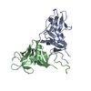
| ||||||||
|---|---|---|---|---|---|---|---|---|---|
| 1 |
| ||||||||
| Unit cell |
|
-Components
| #1: Protein | Mass: 15668.785 Da / Num. of mol.: 2 Source method: isolated from a genetically manipulated source Source: (gene. exp.) Homo sapiens (human) / Gene: OLR1, CLEC8A, LOX1 / Plasmid: pET-15b / Production host:  Escherichia coli B (bacteria) / References: UniProt: P78380 Escherichia coli B (bacteria) / References: UniProt: P78380#2: Water | ChemComp-HOH / | Water Mass: 18.015 Da / Num. of mol.: 369 / Source method: isolated from a natural source / Formula: H2O Mass: 18.015 Da / Num. of mol.: 369 / Source method: isolated from a natural source / Formula: H2O |
|---|
-Experimental details
-Experiment
| Experiment | Method: X-RAY DIFFRACTION / Number of used crystals: 1 |
|---|
- Sample preparation
| Crystal | Density Matthews: 2.21 Å3/Da / Density % sol: 44.24 % |
|---|---|
| Crystal grow | Temperature: 293.15 K / Method: vapor diffusion, sitting drop / pH: 7 Details: protein 8mg/ml, 10mM Tris-HCl, 150mM NaCl, 1.1 M Sodium Malonate, 0.1M HEPES pH 7, 0.5% v/v Jeffamine ED-2001 |
-Data collection
| Diffraction | Mean temperature: 100 K / Serial crystal experiment: N | |||||||||||||||||||||||||||
|---|---|---|---|---|---|---|---|---|---|---|---|---|---|---|---|---|---|---|---|---|---|---|---|---|---|---|---|---|
| Diffraction source | Source: SYNCHROTRON / Site: ESRF  / Beamline: ID29 / Wavelength: 0.984 Å / Beamline: ID29 / Wavelength: 0.984 Å | |||||||||||||||||||||||||||
| Detector | Type: DECTRIS PILATUS 6M-F / Detector: PIXEL / Date: Jul 16, 2017 | |||||||||||||||||||||||||||
| Radiation | Protocol: SINGLE WAVELENGTH / Monochromatic (M) / Laue (L): M / Scattering type: x-ray | |||||||||||||||||||||||||||
| Radiation wavelength | Wavelength: 0.984 Å / Relative weight: 1 | |||||||||||||||||||||||||||
| Reflection | Resolution: 1.01→75.724 Å / Num. obs: 105602 / % possible obs: 91.2 % / Redundancy: 2.9 % / Biso Wilson estimate: 12.03 Å2 / CC1/2: 0.999 / Rmerge(I) obs: 0.037 / Rpim(I) all: 0.025 / Rrim(I) all: 0.045 / Net I/σ(I): 12 / Num. measured all: 307143 | |||||||||||||||||||||||||||
| Reflection shell | Diffraction-ID: 1 / Redundancy: 2.9 %
|
- Processing
| Software |
| ||||||||||||||||||||||||||||||||||||||||||||||||||||||||||||||||||||||||||||||||||||||||||||||||||||||||||||
|---|---|---|---|---|---|---|---|---|---|---|---|---|---|---|---|---|---|---|---|---|---|---|---|---|---|---|---|---|---|---|---|---|---|---|---|---|---|---|---|---|---|---|---|---|---|---|---|---|---|---|---|---|---|---|---|---|---|---|---|---|---|---|---|---|---|---|---|---|---|---|---|---|---|---|---|---|---|---|---|---|---|---|---|---|---|---|---|---|---|---|---|---|---|---|---|---|---|---|---|---|---|---|---|---|---|---|---|---|---|
| Refinement | Method to determine structure: MOLECULAR REPLACEMENT Starting model: 1YPQ Resolution: 1.141→16.62 Å / Cor.coef. Fo:Fc: 0.965 / Cor.coef. Fo:Fc free: 0.959 / SU R Cruickshank DPI: 0.036 / Cross valid method: THROUGHOUT / σ(F): 0 / SU R Blow DPI: 0.038 / SU Rfree Blow DPI: 0.039 / SU Rfree Cruickshank DPI: 0.036
| ||||||||||||||||||||||||||||||||||||||||||||||||||||||||||||||||||||||||||||||||||||||||||||||||||||||||||||
| Displacement parameters | Biso max: 74.77 Å2 / Biso mean: 17.33 Å2 / Biso min: 7.63 Å2
| ||||||||||||||||||||||||||||||||||||||||||||||||||||||||||||||||||||||||||||||||||||||||||||||||||||||||||||
| Refine analyze | Luzzati coordinate error obs: 0.13 Å | ||||||||||||||||||||||||||||||||||||||||||||||||||||||||||||||||||||||||||||||||||||||||||||||||||||||||||||
| Refinement step | Cycle: final / Resolution: 1.141→16.62 Å
| ||||||||||||||||||||||||||||||||||||||||||||||||||||||||||||||||||||||||||||||||||||||||||||||||||||||||||||
| Refine LS restraints |
| ||||||||||||||||||||||||||||||||||||||||||||||||||||||||||||||||||||||||||||||||||||||||||||||||||||||||||||
| LS refinement shell | Resolution: 1.141→1.15 Å / Rfactor Rfree error: 0
| ||||||||||||||||||||||||||||||||||||||||||||||||||||||||||||||||||||||||||||||||||||||||||||||||||||||||||||
| Refinement TLS params. | Method: refined / Refine-ID: X-RAY DIFFRACTION
| ||||||||||||||||||||||||||||||||||||||||||||||||||||||||||||||||||||||||||||||||||||||||||||||||||||||||||||
| Refinement TLS group |
|
