 Movie
Movie Controller


+ Open data

- Basic information
| Entry | Database: PDB / ID: 7w15 | ||||||
|---|---|---|---|---|---|---|---|
| Title | Crystal Structure of MPH-E in complex with GTP and Erythromycin | ||||||
 Components Components | Macrolide 2'-phosphotransferase | ||||||
 Keywords Keywords |  TRANSFERASE / Antibotic resistance TRANSFERASE / Antibotic resistance | ||||||
| Function / homology | Aminoglycoside phosphotransferase / Phosphotransferase enzyme family / transferase activity / GTP binding / Protein kinase-like domain superfamily / metal ion binding / ERYTHROMYCIN A / GUANOSINE-5'-TRIPHOSPHATE / Macrolide 2'-phosphotransferase Function and homology information Function and homology information | ||||||
| Biological species |  Acinetobacter baumannii (bacteria) Acinetobacter baumannii (bacteria) | ||||||
| Method | X-RAY DIFFRACTION / SYNCHROTRON / MOLECULAR REPLACEMENT / Resolution: 1.77 Å | ||||||
 Authors Authors | Qi, Q. / Kuang, L. / Jiang, Y. | ||||||
| Funding support |  China, 1items China, 1items
| ||||||
 Citation Citation | Journal: To Be Published Title: Crystal Structure of the Acinetobacter baumannii Macrolide Phosphotransferases E Reveal the Novel Catalysis Mechanism Authors: Qi, Q. / Kuang, L. / Jiang, Y. | ||||||
| History |
|
- Structure visualization
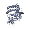
| Structure viewer | Molecule: MolmilJmol/JSmol |
|---|
- Downloads & links
-Download
| PDBx/mmCIF format | 7w15.cif.gz | 345.1 KB | Display | PDBx/mmCIF format |
|---|---|---|---|---|
| PDB format | pdb7w15.ent.gz | 232.5 KB | Display | PDB format |
| PDBx/mmJSON format | 7w15.json.gz | Tree view | PDBx/mmJSON format | |
| Others |  Other downloads Other downloads |
-Validation report
| Arichive directory | https://data.pdbj.org/pub/pdb/validation_reports/w1/7w15ftp://data.pdbj.org/pub/pdb/validation_reports/w1/7w15 | HTTPS FTP |
|---|
-Related structure data
-Links
 PDBj
PDBj


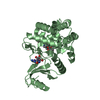
- Assembly
| Deposited unit | 
| |||||||||||||||||||||||||||||||||||||||||||||||||||||||||||||||||||||||||||||||
|---|---|---|---|---|---|---|---|---|---|---|---|---|---|---|---|---|---|---|---|---|---|---|---|---|---|---|---|---|---|---|---|---|---|---|---|---|---|---|---|---|---|---|---|---|---|---|---|---|---|---|---|---|---|---|---|---|---|---|---|---|---|---|---|---|---|---|---|---|---|---|---|---|---|---|---|---|---|---|---|---|
| 1 | 
| |||||||||||||||||||||||||||||||||||||||||||||||||||||||||||||||||||||||||||||||
| 2 | 
| |||||||||||||||||||||||||||||||||||||||||||||||||||||||||||||||||||||||||||||||
| Unit cell |
| |||||||||||||||||||||||||||||||||||||||||||||||||||||||||||||||||||||||||||||||
| Noncrystallographic symmetry (NCS) | NCS domain:
NCS domain segments: Ens-ID: ens_1
|
-Components
-Protein , 1 types, 2 molecules BA
| #1: Protein | Mass: 34619.586 Da / Num. of mol.: 2 Source method: isolated from a genetically manipulated source Source: (gene. exp.) Acinetobacter baumannii (bacteria) / Gene: mph(E)Production host: Escherichia coli 'BL21-Gold(DE3)pLysS AG' (bacteria)References: UniProt: A5Y459 |
|---|
-Non-polymers , 5 types, 845 molecules 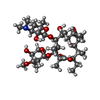


| #2: Chemical | Erythromycin Mass: 733.927 Da / Num. of mol.: 2 / Source method: obtained synthetically / Formula: C37H67NO13 / Feature type: SUBJECT OF INVESTIGATION / Comment: antibiotic*YM Mass: 733.927 Da / Num. of mol.: 2 / Source method: obtained synthetically / Formula: C37H67NO13 / Feature type: SUBJECT OF INVESTIGATION / Comment: antibiotic*YM#3: Chemical | Guanosine triphosphate Mass: 523.180 Da / Num. of mol.: 2 / Source method: obtained synthetically / Formula: C10H16N5O14P3 / Feature type: SUBJECT OF INVESTIGATION / Comment: GTP, energy-carrying molecule*YM Mass: 523.180 Da / Num. of mol.: 2 / Source method: obtained synthetically / Formula: C10H16N5O14P3 / Feature type: SUBJECT OF INVESTIGATION / Comment: GTP, energy-carrying molecule*YM#4: Chemical | ChemComp-MG /  Mass: 24.305 Da / Num. of mol.: 6 / Source method: obtained synthetically / Formula: Mg / Feature type: SUBJECT OF INVESTIGATION Mass: 24.305 Da / Num. of mol.: 6 / Source method: obtained synthetically / Formula: Mg / Feature type: SUBJECT OF INVESTIGATION#5: Chemical | Glycerol Mass: 92.094 Da / Num. of mol.: 2 / Source method: obtained synthetically / Formula: C3H8O3 Mass: 92.094 Da / Num. of mol.: 2 / Source method: obtained synthetically / Formula: C3H8O3#6: Water | ChemComp-HOH / | WaterMass: 18.015 Da / Num. of mol.: 833 / Source method: isolated from a natural source / Formula: H2O |
|---|
-Details
| Has ligand of interest | Y |
|---|
-Experimental details
-Experiment
| Experiment | Method: X-RAY DIFFRACTION / Number of used crystals: 1 |
|---|
- Sample preparation
| Crystal | Density Matthews: 2.7 Å3/Da / Density % sol: 54.49 % |
|---|---|
| Crystal grow | Temperature: 293.15 K / Method: vapor diffusion, sitting drop Details: O.O8 M Mggnesium acetate tetrahydrate; 0.05 M sodium cacodylate pH6.5; 25% PEG3350 |
-Data collection
| Diffraction | Mean temperature: 100 K / Serial crystal experiment: N |
|---|---|
| Diffraction source | Source: SYNCHROTRON / Site: SSRF / Beamline: BL18U1 / Wavelength: 0.973 Å |
| Detector | Type: DECTRIS PILATUS3 6M / Detector: PIXEL / Date: May 30, 2021 |
| Radiation | Protocol: SINGLE WAVELENGTH / Monochromatic (M) / Laue (L): M / Scattering type: x-ray |
| Radiation wavelength | Wavelength: 0.973 Å / Relative weight: 1 |
| Reflection | Resolution: 1.77→50 Å / Num. obs: 72216 / % possible obs: 99.7 % / Redundancy: 6.7 % / Biso Wilson estimate: 19.92 Å2 / CC1/2: 0.988 / Rrim(I) all: 0.107 / Net I/σ(I): 19.7 |
| Reflection shell | Resolution: 1.77→1.8 Å / Mean I/σ(I) obs: 3 / Num. unique obs: 3600 / CC1/2: 0.872 / Rrim(I) all: 0.517 / % possible all: 96.5 |
- Processing
| Software |
| |||||||||||||||||||||||||||||||||||||||||||||||||||||||||||||||||||||||||||||||||||||||||||||||||||||||||
|---|---|---|---|---|---|---|---|---|---|---|---|---|---|---|---|---|---|---|---|---|---|---|---|---|---|---|---|---|---|---|---|---|---|---|---|---|---|---|---|---|---|---|---|---|---|---|---|---|---|---|---|---|---|---|---|---|---|---|---|---|---|---|---|---|---|---|---|---|---|---|---|---|---|---|---|---|---|---|---|---|---|---|---|---|---|---|---|---|---|---|---|---|---|---|---|---|---|---|---|---|---|---|---|---|---|---|
| Refinement | Method to determine structure: MOLECULAR REPLACEMENT / Resolution: 1.77→34.85 Å / SU ML: 0.1681 / Cross valid method: FREE R-VALUE / σ(F): 1.38 / Phase error: 19.1932 Stereochemistry target values: GeoStd + Monomer Library + CDL v1.2
| |||||||||||||||||||||||||||||||||||||||||||||||||||||||||||||||||||||||||||||||||||||||||||||||||||||||||
| Solvent computation | Shrinkage radii: 0.9 Å / VDW probe radii: 1.1 Å / Solvent model: FLAT BULK SOLVENT MODEL | |||||||||||||||||||||||||||||||||||||||||||||||||||||||||||||||||||||||||||||||||||||||||||||||||||||||||
| Displacement parameters | Biso mean: 26.23 Å2 | |||||||||||||||||||||||||||||||||||||||||||||||||||||||||||||||||||||||||||||||||||||||||||||||||||||||||
| Refinement step | Cycle: LAST / Resolution: 1.77→34.85 Å
| |||||||||||||||||||||||||||||||||||||||||||||||||||||||||||||||||||||||||||||||||||||||||||||||||||||||||
| Refine LS restraints |
| |||||||||||||||||||||||||||||||||||||||||||||||||||||||||||||||||||||||||||||||||||||||||||||||||||||||||
| Refine LS restraints NCS | Type: Torsion NCS / Rms dev position: 0.599001571713 Å | |||||||||||||||||||||||||||||||||||||||||||||||||||||||||||||||||||||||||||||||||||||||||||||||||||||||||
| LS refinement shell |
| |||||||||||||||||||||||||||||||||||||||||||||||||||||||||||||||||||||||||||||||||||||||||||||||||||||||||
| Refinement TLS params. | Method: refined / Origin x: -1.93796165461 Å / Origin y: -13.3804685763 Å / Origin z: 24.8057055858 Å
| |||||||||||||||||||||||||||||||||||||||||||||||||||||||||||||||||||||||||||||||||||||||||||||||||||||||||
| Refinement TLS group | Selection details: all |
