 Movie
Movie Controller


+ Open data

- Basic information
| Entry | Database: PDB / ID: 7w0t | ||||||
|---|---|---|---|---|---|---|---|
| Title | TRIM7 in complex with C-terminal peptide of 2C | ||||||
 Components Components |
| ||||||
 Keywords Keywords |  ANTIVIRAL PROTEIN ANTIVIRAL PROTEIN | ||||||
| Function / homology |  Function and homology information Function and homology informationantiviral innate immune response / RING-type E3 ubiquitin transferase / ubiquitin protein ligase activity / protein ubiquitination / Golgi apparatus / zinc ion binding / nucleus / cytoplasmSimilarity search - Function | ||||||
| Biological species |  Homo sapiens (human) Homo sapiens (human) Coxsackievirus Coxsackievirus | ||||||
| Method | X-RAY DIFFRACTION / SYNCHROTRON / MOLECULAR REPLACEMENT / Resolution: 1.57 Å | ||||||
 Authors Authors | Zhang, H. / Liang, X. / Li, X.Z. | ||||||
| Funding support | 1items
| ||||||
 Citation Citation | Journal: Nat.Chem.Biol. / Year: 2022 Title: A C-terminal glutamine recognition mechanism revealed by E3 ligase TRIM7 structures. Authors: Liang, X. / Xiao, J. / Li, X. / Liu, Y. / Lu, Y. / Wen, Y. / Li, Z. / Che, X. / Ma, Y. / Zhang, X. / Zhang, Y. / Jian, D. / Wang, P. / Xuan, C. / Yu, G. / Li, L. / Zhang, H. | ||||||
| History |
|
- Structure visualization
| Structure viewer | Molecule: MolmilJmol/JSmol |
|---|
- Downloads & links
-Download
| PDBx/mmCIF format | 7w0t.cif.gz | 272.4 KB | Display | PDBx/mmCIF format |
|---|---|---|---|---|
| PDB format | pdb7w0t.ent.gz | 179.5 KB | Display | PDB format |
| PDBx/mmJSON format | 7w0t.json.gz | Tree view | PDBx/mmJSON format | |
| Others |  Other downloads Other downloads |
-Validation report
| Arichive directory | https://data.pdbj.org/pub/pdb/validation_reports/w0/7w0tftp://data.pdbj.org/pub/pdb/validation_reports/w0/7w0t | HTTPS FTP |
|---|
-Related structure data
| Related structure data | 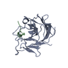 7w0qC 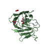 7w0sC  7x6yC 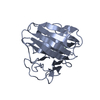 7x6zC  7x70C 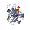 6umaS S: Starting model for refinement C: citing same article ( |
|---|---|
| Similar structure data |
-Links
 PDBj
PDBj

- Assembly
| Deposited unit | 
| ||||||||||
|---|---|---|---|---|---|---|---|---|---|---|---|
| 1 | 
| ||||||||||
| 2 | 
| ||||||||||
| 3 | 
| ||||||||||
| Unit cell |
|
-Components
| #1: Protein | Mass: 19689.295 Da / Num. of mol.: 3 Source method: isolated from a genetically manipulated source Source: (gene. exp.) Homo sapiens (human) / Gene: TRIM7, GNIP, RNF90 / Production host:  Escherichia coli (E. coli) Escherichia coli (E. coli)References: UniProt: Q9C029, RING-type E3 ubiquitin transferase #2: Protein/peptide | Mass: 1078.215 Da / Num. of mol.: 3 / Source method: obtained synthetically / Source: (synth.) Coxsackievirus#3: Water | ChemComp-HOH / | Water Mass: 18.015 Da / Num. of mol.: 625 / Source method: isolated from a natural source / Formula: H2O Mass: 18.015 Da / Num. of mol.: 625 / Source method: isolated from a natural source / Formula: H2O |
|---|
-Experimental details
-Experiment
| Experiment | Method: X-RAY DIFFRACTION / Number of used crystals: 1 |
|---|
- Sample preparation
| Crystal | Density Matthews: 2.43 Å3/Da / Density % sol: 49.48 % |
|---|---|
| Crystal grow | Temperature: 291.15 K / Method: liquid diffusion Details: ammonium acetate, magnesium acetate tetrahydrate, HEPES sodium, polyethylene glycol |
-Data collection
| Diffraction | Mean temperature: 100 K / Serial crystal experiment: N |
|---|---|
| Diffraction source | Source: SYNCHROTRON / Site: SSRF  / Beamline: BL18U1 / Wavelength: 0.979 Å / Beamline: BL18U1 / Wavelength: 0.979 Å |
| Detector | Type: DECTRIS PILATUS3 6M / Detector: PIXEL / Date: Oct 6, 2021 |
| Radiation | Protocol: SINGLE WAVELENGTH / Monochromatic (M) / Laue (L): M / Scattering type: x-ray |
| Radiation wavelength | Wavelength: 0.979 Å / Relative weight: 1 |
| Reflection | Resolution: 1.569→42.538 Å / Num. obs: 83192 / % possible obs: 99.03 % / Redundancy: 6.7 % / Biso Wilson estimate: 17.96 Å2 / CC1/2: 0.999 / Net I/σ(I): 14.33 |
| Reflection shell | Resolution: 1.569→1.625 Å / Num. unique obs: 8288 / CC1/2: 0.776 |
- Processing
| Software |
| |||||||||||||||||||||||||||||||||||||||||||||||||||||||||||||||||||||||||||||||||||||||||||||||||||||||||||||||||||||||||||||||||||||||||||||||||||||||||||||||||||||||||||||||||||||||||||||||||||||||||||||||||||||||||
|---|---|---|---|---|---|---|---|---|---|---|---|---|---|---|---|---|---|---|---|---|---|---|---|---|---|---|---|---|---|---|---|---|---|---|---|---|---|---|---|---|---|---|---|---|---|---|---|---|---|---|---|---|---|---|---|---|---|---|---|---|---|---|---|---|---|---|---|---|---|---|---|---|---|---|---|---|---|---|---|---|---|---|---|---|---|---|---|---|---|---|---|---|---|---|---|---|---|---|---|---|---|---|---|---|---|---|---|---|---|---|---|---|---|---|---|---|---|---|---|---|---|---|---|---|---|---|---|---|---|---|---|---|---|---|---|---|---|---|---|---|---|---|---|---|---|---|---|---|---|---|---|---|---|---|---|---|---|---|---|---|---|---|---|---|---|---|---|---|---|---|---|---|---|---|---|---|---|---|---|---|---|---|---|---|---|---|---|---|---|---|---|---|---|---|---|---|---|---|---|---|---|---|---|---|---|---|---|---|---|---|---|---|---|---|---|---|---|---|
| Refinement | Method to determine structure: MOLECULAR REPLACEMENT Starting model: 6UMA Resolution: 1.57→42.45 Å / SU ML: 0.1632 / Cross valid method: FREE R-VALUE / σ(F): 1.35 / Phase error: 18.5949 Stereochemistry target values: GeoStd + Monomer Library + CDL v1.2
| |||||||||||||||||||||||||||||||||||||||||||||||||||||||||||||||||||||||||||||||||||||||||||||||||||||||||||||||||||||||||||||||||||||||||||||||||||||||||||||||||||||||||||||||||||||||||||||||||||||||||||||||||||||||||
| Solvent computation | Shrinkage radii: 0.9 Å / VDW probe radii: 1.11 Å / Solvent model: FLAT BULK SOLVENT MODEL | |||||||||||||||||||||||||||||||||||||||||||||||||||||||||||||||||||||||||||||||||||||||||||||||||||||||||||||||||||||||||||||||||||||||||||||||||||||||||||||||||||||||||||||||||||||||||||||||||||||||||||||||||||||||||
| Displacement parameters | Biso mean: 21.77 Å2 | |||||||||||||||||||||||||||||||||||||||||||||||||||||||||||||||||||||||||||||||||||||||||||||||||||||||||||||||||||||||||||||||||||||||||||||||||||||||||||||||||||||||||||||||||||||||||||||||||||||||||||||||||||||||||
| Refinement step | Cycle: LAST / Resolution: 1.57→42.45 Å
| |||||||||||||||||||||||||||||||||||||||||||||||||||||||||||||||||||||||||||||||||||||||||||||||||||||||||||||||||||||||||||||||||||||||||||||||||||||||||||||||||||||||||||||||||||||||||||||||||||||||||||||||||||||||||
| Refine LS restraints |
| |||||||||||||||||||||||||||||||||||||||||||||||||||||||||||||||||||||||||||||||||||||||||||||||||||||||||||||||||||||||||||||||||||||||||||||||||||||||||||||||||||||||||||||||||||||||||||||||||||||||||||||||||||||||||
| LS refinement shell |
| |||||||||||||||||||||||||||||||||||||||||||||||||||||||||||||||||||||||||||||||||||||||||||||||||||||||||||||||||||||||||||||||||||||||||||||||||||||||||||||||||||||||||||||||||||||||||||||||||||||||||||||||||||||||||
| Refinement TLS params. | Method: refined / Origin x: -32.9975494094 Å / Origin y: -9.46656087141 Å / Origin z: 20.4040875467 Å
| |||||||||||||||||||||||||||||||||||||||||||||||||||||||||||||||||||||||||||||||||||||||||||||||||||||||||||||||||||||||||||||||||||||||||||||||||||||||||||||||||||||||||||||||||||||||||||||||||||||||||||||||||||||||||
| Refinement TLS group | Selection details: all |
