 Movie
Movie Controller

[English] 日本語
 Yorodumi
- PDB-7vwp: Structure of the flavin-dependent monooxygenase FlsO1 from the bi... -
+ Open data

- Basic information
| Entry | Database: PDB / ID: 7vwp | ||||||
|---|---|---|---|---|---|---|---|
| Title | Structure of the flavin-dependent monooxygenase FlsO1 from the biosynthesis of fluostatinsin | ||||||
 Components Components | FlsO1 | ||||||
 Keywords Keywords |  OXIDOREDUCTASE / hydroxylation / benzo[b]-fluorene / epoxidation / Baeyer-Villiger Oxidation / prejadomycin OXIDOREDUCTASE / hydroxylation / benzo[b]-fluorene / epoxidation / Baeyer-Villiger Oxidation / prejadomycin | ||||||
| Function / homology | FAD-binding domain / FAD binding domain / FAD binding / FAD/NAD(P)-binding domain superfamily / FLAVIN-ADENINE DINUCLEOTIDE / PHOSPHATE ION / FlsO1 Function and homology information Function and homology information | ||||||
| Biological species |  Micromonospora rosaria (bacteria) Micromonospora rosaria (bacteria) | ||||||
| Method | X-RAY DIFFRACTION / MOLECULAR REPLACEMENT / Resolution: 2.3 Å | ||||||
 Authors Authors | Zhang, Y. / Yang, C. / Zhang, L. / Zhang, C. | ||||||
| Funding support |  China, 1items China, 1items
| ||||||
 Citation Citation | Journal: Nat Commun / Year: 2022 Title: Biochemical and structural insights of multifunctional flavin-dependent monooxygenase FlsO1-catalyzed unexpected xanthone formation Authors: Yang, C. / Zhang, L. / Zhang, W. / Huang, C. / Zhu, Y. / Jiang, X. / Liu, W. / Zhao, M. / De, B.C. / Zhang, C. | ||||||
| History |
|
- Structure visualization
| Structure viewer | Molecule: MolmilJmol/JSmol |
|---|
- Downloads & links
-Download
| PDBx/mmCIF format | 7vwp.cif.gz | 407.8 KB | Display | PDBx/mmCIF format |
|---|---|---|---|---|
| PDB format | pdb7vwp.ent.gz | 318.7 KB | Display | PDB format |
| PDBx/mmJSON format | 7vwp.json.gz | Tree view | PDBx/mmJSON format | |
| Others |  Other downloads Other downloads |
-Validation report
| Arichive directory | https://data.pdbj.org/pub/pdb/validation_reports/vw/7vwpftp://data.pdbj.org/pub/pdb/validation_reports/vw/7vwp | HTTPS FTP |
|---|
-Related structure data
| Related structure data | 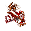 2qa1S S: Starting model for refinement |
|---|---|
| Similar structure data |
-Links
 PDBj
PDBj- Assembly
| Deposited unit | 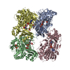
| ||||||||
|---|---|---|---|---|---|---|---|---|---|
| 1 | 
| ||||||||
| 2 | 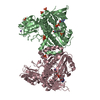
| ||||||||
| Unit cell |
|
-Components
| #1: Protein | Mass: 53276.980 Da / Num. of mol.: 4 Source method: isolated from a genetically manipulated source Source: (gene. exp.) Micromonospora rosaria (bacteria) / Production host: Escherichia coli BL21(DE3) (bacteria) / Strain (production host): BL21(DE3) / References: UniProt: A0A0P0I576#2: Chemical | ChemComp-PO4 / Phosphate  Mass: 94.971 Da / Num. of mol.: 14 / Source method: obtained synthetically / Formula: PO4 Mass: 94.971 Da / Num. of mol.: 14 / Source method: obtained synthetically / Formula: PO4#3: Chemical | ChemComp-FAD / Flavin adenine dinucleotide  Mass: 785.550 Da / Num. of mol.: 4 / Source method: obtained synthetically / Formula: C27H33N9O15P2 / Feature type: SUBJECT OF INVESTIGATION / Comment: FAD*YM Mass: 785.550 Da / Num. of mol.: 4 / Source method: obtained synthetically / Formula: C27H33N9O15P2 / Feature type: SUBJECT OF INVESTIGATION / Comment: FAD*YM#4: Chemical | ChemComp-NA / |   Mass: 22.990 Da / Num. of mol.: 1 / Source method: obtained synthetically / Formula: Na Mass: 22.990 Da / Num. of mol.: 1 / Source method: obtained synthetically / Formula: Na#5: Water | ChemComp-HOH / | Water Mass: 18.015 Da / Num. of mol.: 1015 / Source method: isolated from a natural source / Formula: H2O Mass: 18.015 Da / Num. of mol.: 1015 / Source method: isolated from a natural source / Formula: H2OHas ligand of interest | Y | |
|---|
-Experimental details
-Experiment
| Experiment | Method: X-RAY DIFFRACTION / Number of used crystals: 1 |
|---|
- Sample preparation
| Crystal | Density Matthews: 2.64 Å3/Da / Density % sol: 53.47 % |
|---|---|
| Crystal grow | Temperature: 289 K / Method: vapor diffusion, sitting drop / pH: 6.5 Details: 0.02 M Sodium/potassium phosphate, 0.1 M Bis-Tris propane, pH 6.5, 20 % w/v PEG 3350 |
-Data collection
| Diffraction | Mean temperature: 100 K / Serial crystal experiment: N |
|---|---|
| Diffraction source | Source: ROTATING ANODE / Type: RIGAKU / Wavelength: 1.54184 Å |
| Detector | Type: DECTRIS PILATUS 200K / Detector: PIXEL / Date: Mar 6, 2019 |
| Radiation | Protocol: SINGLE WAVELENGTH / Monochromatic (M) / Laue (L): M / Scattering type: x-ray |
| Radiation wavelength | Wavelength: 1.54184 Å / Relative weight: 1 |
| Reflection | Resolution: 2.3→13.182 Å / Num. obs: 95967 / % possible obs: 98.9 % / Redundancy: 3.6 % / Rmerge(I) obs: 0.066 / Net I/σ(I): 12.9 |
| Reflection shell | Resolution: 2.3→2.34 Å / Rmerge(I) obs: 0.39 / Mean I/σ(I) obs: 2.8 / Num. unique obs: 4703 |
- Processing
| Software |
| ||||||||||||||||||||||||||||||||||||||||||||||||||||||||||||||||||||||||||||||||||||||||||||||||||||||||||||||||||||||||||||||||||||||||||||||||||||||||||||||||
|---|---|---|---|---|---|---|---|---|---|---|---|---|---|---|---|---|---|---|---|---|---|---|---|---|---|---|---|---|---|---|---|---|---|---|---|---|---|---|---|---|---|---|---|---|---|---|---|---|---|---|---|---|---|---|---|---|---|---|---|---|---|---|---|---|---|---|---|---|---|---|---|---|---|---|---|---|---|---|---|---|---|---|---|---|---|---|---|---|---|---|---|---|---|---|---|---|---|---|---|---|---|---|---|---|---|---|---|---|---|---|---|---|---|---|---|---|---|---|---|---|---|---|---|---|---|---|---|---|---|---|---|---|---|---|---|---|---|---|---|---|---|---|---|---|---|---|---|---|---|---|---|---|---|---|---|---|---|---|---|---|---|
| Refinement | Method to determine structure: MOLECULAR REPLACEMENT Starting model: 2QA1 Resolution: 2.3→13.182 Å / Cor.coef. Fo:Fc: 0.938 / Cor.coef. Fo:Fc free: 0.893 / SU B: 8.819 / SU ML: 0.207 / Cross valid method: FREE R-VALUE / ESU R: 0.374 / ESU R Free: 0.265 Details: Hydrogens have been added in their riding positions
| ||||||||||||||||||||||||||||||||||||||||||||||||||||||||||||||||||||||||||||||||||||||||||||||||||||||||||||||||||||||||||||||||||||||||||||||||||||||||||||||||
| Solvent computation | Ion probe radii: 0.8 Å / Shrinkage radii: 0.8 Å / VDW probe radii: 1.2 Å / Solvent model: MASK BULK SOLVENT | ||||||||||||||||||||||||||||||||||||||||||||||||||||||||||||||||||||||||||||||||||||||||||||||||||||||||||||||||||||||||||||||||||||||||||||||||||||||||||||||||
| Displacement parameters | Biso mean: 29.263 Å2
| ||||||||||||||||||||||||||||||||||||||||||||||||||||||||||||||||||||||||||||||||||||||||||||||||||||||||||||||||||||||||||||||||||||||||||||||||||||||||||||||||
| Refinement step | Cycle: LAST / Resolution: 2.3→13.182 Å
| ||||||||||||||||||||||||||||||||||||||||||||||||||||||||||||||||||||||||||||||||||||||||||||||||||||||||||||||||||||||||||||||||||||||||||||||||||||||||||||||||
| Refine LS restraints |
| ||||||||||||||||||||||||||||||||||||||||||||||||||||||||||||||||||||||||||||||||||||||||||||||||||||||||||||||||||||||||||||||||||||||||||||||||||||||||||||||||
| LS refinement shell |
|
