 Movie
Movie Controller


+ Open data

- Basic information
| Entry | Database: PDB / ID: 7vwn | |||||||||
|---|---|---|---|---|---|---|---|---|---|---|
| Title | The structure of an engineered PET hydrolase | |||||||||
 Components Components | Poly(ethylene terephthalate) hydrolase | |||||||||
 Keywords Keywords |  HYDROLASE / PET hydrolase / PETase / biodegradation of microplastics HYDROLASE / PET hydrolase / PETase / biodegradation of microplastics | |||||||||
| Function / homology |  Function and homology information Function and homology informationpoly(ethylene terephthalate) hydrolase / acetylesterase activity / carboxylic ester hydrolase activity / cellular response to organic substance / xenobiotic catabolic process / extracellular regionSimilarity search - Function | |||||||||
| Biological species |  Ideonella sakaiensis (bacteria) Ideonella sakaiensis (bacteria) | |||||||||
| Method | X-RAY DIFFRACTION / MOLECULAR REPLACEMENT / Resolution: 1.45 Å | |||||||||
 Authors Authors | Xie, W. / Jia, Q. | |||||||||
| Funding support |  China, 2items China, 2items
| |||||||||
 Citation Citation | Journal: To Be Published Title: An engineered PET hydrolase for biodegradation of microplastics in ocean water Authors: Xie, W. / Jia, Q. | |||||||||
| History |
|
- Structure visualization
| Structure viewer | Molecule: MolmilJmol/JSmol |
|---|
- Downloads & links
-Download
| PDBx/mmCIF format | 7vwn.cif.gz | 147.2 KB | Display | PDBx/mmCIF format |
|---|---|---|---|---|
| PDB format | pdb7vwn.ent.gz | 94.2 KB | Display | PDB format |
| PDBx/mmJSON format | 7vwn.json.gz | Tree view | PDBx/mmJSON format | |
| Others |  Other downloads Other downloads |
-Validation report
| Arichive directory | https://data.pdbj.org/pub/pdb/validation_reports/vw/7vwnftp://data.pdbj.org/pub/pdb/validation_reports/vw/7vwn | HTTPS FTP |
|---|
-Related structure data
| Related structure data | 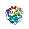 6eqeS S: Starting model for refinement |
|---|---|
| Similar structure data |
-Links
 PDBj
PDBj
- Assembly
| Deposited unit | 
| ||||||||||||
|---|---|---|---|---|---|---|---|---|---|---|---|---|---|
| 1 |
| ||||||||||||
| Unit cell |
| ||||||||||||
| Components on special symmetry positions |
|
-Components
| #1: Protein | Mass: 28515.600 Da / Num. of mol.: 1 Mutation: T29S, K95N, I168R, P181V, S214V, N233C, A248D, R280A, S282C Source method: isolated from a genetically manipulated source Source: (gene. exp.) Ideonella sakaiensis (strain NBRC 110686 / TISTR 2288 / 201-F6) (bacteria)Strain: NBRC 110686 / TISTR 2288 / 201-F6 / Gene: ISF6_4831 / Production host: Escherichia coli (E. coli)References: UniProt: A0A0K8P6T7, poly(ethylene terephthalate) hydrolase | ||||
|---|---|---|---|---|---|
| #2: Chemical | Chloride  Mass: 35.453 Da / Num. of mol.: 3 / Source method: obtained synthetically / Formula: Cl / Feature type: SUBJECT OF INVESTIGATION Mass: 35.453 Da / Num. of mol.: 3 / Source method: obtained synthetically / Formula: Cl / Feature type: SUBJECT OF INVESTIGATION#3: Water | ChemComp-HOH / | Water Mass: 18.015 Da / Num. of mol.: 282 / Source method: isolated from a natural source / Formula: H2O Mass: 18.015 Da / Num. of mol.: 282 / Source method: isolated from a natural source / Formula: H2OHas ligand of interest | Y | |
-Experimental details
-Experiment
| Experiment | Method: X-RAY DIFFRACTION / Number of used crystals: 1 |
|---|
- Sample preparation
| Crystal | Density Matthews: 2.12 Å3/Da / Density % sol: 41.86 % |
|---|---|
| Crystal grow | Temperature: 298 K / Method: vapor diffusion, sitting drop / pH: 6 / Details: 1.8 M (NH4)2SO4, 0.1 M NaCl and 0.1 M MES pH6.0 |
-Data collection
| Diffraction | Mean temperature: 100 K / Serial crystal experiment: N |
|---|---|
| Diffraction source | Source: ROTATING ANODE / Type: RIGAKU MICROMAX-007 HF / Wavelength: 0.979 Å |
| Detector | Type: OXFORD ONYX CCD / Detector: CCD / Date: Jul 7, 2021 |
| Radiation | Protocol: SINGLE WAVELENGTH / Monochromatic (M) / Laue (L): M / Scattering type: x-ray |
| Radiation wavelength | Wavelength: 0.979 Å / Relative weight: 1 |
| Reflection | Resolution: 1.45→50 Å / Num. obs: 42326 / % possible obs: 98.2 % / Redundancy: 6.7 % / Biso Wilson estimate: 15.37 Å2 / CC1/2: 0.99 / Rmerge(I) obs: 0.063 / Net I/σ(I): 23.3 |
| Reflection shell | Resolution: 1.45→1.5 Å / Redundancy: 6 % / Rmerge(I) obs: 0.38 / Mean I/σ(I) obs: 2.8 / Num. unique obs: 3822 / CC1/2: 0.93 / % possible all: 91 |
- Processing
| Software |
| ||||||||||||||||||||||||||||||||||||||||||||||||||||||||||||||||||||||||||||||||||||||||||||||||||||||||||||||||
|---|---|---|---|---|---|---|---|---|---|---|---|---|---|---|---|---|---|---|---|---|---|---|---|---|---|---|---|---|---|---|---|---|---|---|---|---|---|---|---|---|---|---|---|---|---|---|---|---|---|---|---|---|---|---|---|---|---|---|---|---|---|---|---|---|---|---|---|---|---|---|---|---|---|---|---|---|---|---|---|---|---|---|---|---|---|---|---|---|---|---|---|---|---|---|---|---|---|---|---|---|---|---|---|---|---|---|---|---|---|---|---|---|---|
| Refinement | Method to determine structure: MOLECULAR REPLACEMENT Starting model: 6EQE Resolution: 1.45→32.95 Å / SU ML: 0.1257 / Cross valid method: FREE R-VALUE / σ(F): 1.38 / Phase error: 18.4513 / Stereochemistry target values: CDL v1.2
| ||||||||||||||||||||||||||||||||||||||||||||||||||||||||||||||||||||||||||||||||||||||||||||||||||||||||||||||||
| Solvent computation | Shrinkage radii: 0.9 Å / VDW probe radii: 1.11 Å / Solvent model: FLAT BULK SOLVENT MODEL | ||||||||||||||||||||||||||||||||||||||||||||||||||||||||||||||||||||||||||||||||||||||||||||||||||||||||||||||||
| Displacement parameters | Biso mean: 23.81 Å2 | ||||||||||||||||||||||||||||||||||||||||||||||||||||||||||||||||||||||||||||||||||||||||||||||||||||||||||||||||
| Refinement step | Cycle: LAST / Resolution: 1.45→32.95 Å
| ||||||||||||||||||||||||||||||||||||||||||||||||||||||||||||||||||||||||||||||||||||||||||||||||||||||||||||||||
| Refine LS restraints |
| ||||||||||||||||||||||||||||||||||||||||||||||||||||||||||||||||||||||||||||||||||||||||||||||||||||||||||||||||
| LS refinement shell |
| ||||||||||||||||||||||||||||||||||||||||||||||||||||||||||||||||||||||||||||||||||||||||||||||||||||||||||||||||
| Refinement TLS params. | Method: refined / Origin x: 17.3142520429 Å / Origin y: 0.0269757496878 Å / Origin z: 11.5592270117 Å
| ||||||||||||||||||||||||||||||||||||||||||||||||||||||||||||||||||||||||||||||||||||||||||||||||||||||||||||||||
| Refinement TLS group | Selection details: all |
