 Movie
Movie Controller

[English] 日本語
 Yorodumi
- PDB-7vvd: Crystal Structure of the Kv7.1 C-terminal Domain in Complex with ... -
+ Open data

- Basic information
| Entry | Database: PDB / ID: 7vvd | ||||||
|---|---|---|---|---|---|---|---|
| Title | Crystal Structure of the Kv7.1 C-terminal Domain in Complex with Calmodulin disease mutation Q135P | ||||||
 Components Components |
| ||||||
 Keywords Keywords |  SIGNALING PROTEIN/METAL BINDING PROTEIN / KCNQ1 / CaM / SIGNALING PROTEIN / SIGNALING PROTEIN-METAL BINDING PROTEIN complex SIGNALING PROTEIN/METAL BINDING PROTEIN / KCNQ1 / CaM / SIGNALING PROTEIN / SIGNALING PROTEIN-METAL BINDING PROTEIN complex | ||||||
| Function / homology |  Function and homology information Function and homology informationgastrin-induced gastric acid secretion / corticosterone secretion / voltage-gated potassium channel activity involved in atrial cardiac muscle cell action potential repolarization / basolateral part of cell / lumenal side of membrane / negative regulation of voltage-gated potassium channel activity / rhythmic behavior / regulation of gastric acid secretion / stomach development / membrane repolarization during atrial cardiac muscle cell action potential ...gastrin-induced gastric acid secretion / corticosterone secretion / voltage-gated potassium channel activity involved in atrial cardiac muscle cell action potential repolarization / basolateral part of cell / lumenal side of membrane / negative regulation of voltage-gated potassium channel activity / rhythmic behavior / regulation of gastric acid secretion / stomach development / membrane repolarization during atrial cardiac muscle cell action potential / Phase 3 - rapid repolarisation / voltage-gated potassium channel activity involved in cardiac muscle cell action potential repolarization / membrane repolarization during action potential / membrane repolarization during ventricular cardiac muscle cell action potential / regulation of atrial cardiac muscle cell membrane repolarization / iodide transport / Phase 2 - plateau phase / potassium ion export across plasma membrane / membrane repolarization during cardiac muscle cell action potential / intracellular chloride ion homeostasis / renal sodium ion absorption / negative regulation of delayed rectifier potassium channel activity / voltage-gated potassium channel activity involved in ventricular cardiac muscle cell action potential repolarization / atrial cardiac muscle cell action potential / auditory receptor cell development / detection of mechanical stimulus involved in sensory perception of sound / regulation of membrane repolarization / delayed rectifier potassium channel activity / protein phosphatase 1 binding / positive regulation of potassium ion transmembrane transport / Voltage gated Potassium channels / outward rectifier potassium channel activity / potassium ion homeostasis / ventricular cardiac muscle cell action potential / non-motile cilium assembly / regulation of ventricular cardiac muscle cell membrane repolarization / CaM pathway / Cam-PDE 1 activation / intestinal absorption / Sodium/Calcium exchangers / regulation of heart contraction / Calmodulin induced events / Reduction of cytosolic Ca++ levels / CREB1 phosphorylation through the activation of CaMKII/CaMKK/CaMKIV cascasde / Activation of Ca-permeable Kainate Receptor / Loss of phosphorylation of MECP2 at T308 / CREB1 phosphorylation through the activation of Adenylate Cyclase / PKA activation / negative regulation of high voltage-gated calcium channel activity / monoatomic ion channel complex / ciliary base / CaMK IV-mediated phosphorylation of CREB / inner ear morphogenesis / Glycogen breakdown (glycogenolysis) / organelle localization by membrane tethering / negative regulation of calcium ion export across plasma membrane / Activation of RAC1 downstream of NMDARs / mitochondrion-endoplasmic reticulum membrane tethering / CLEC7A (Dectin-1) induces NFAT activation / regulation of cardiac muscle cell action potential / autophagosome membrane docking / positive regulation of heart rate / cochlea development / renal absorption / adrenergic receptor signaling pathway / potassium ion import across plasma membrane / positive regulation of ryanodine-sensitive calcium-release channel activity / Negative regulation of NMDA receptor-mediated neuronal transmission / regulation of cell communication by electrical coupling involved in cardiac conduction / negative regulation of peptidyl-threonine phosphorylation / Unblocking of NMDA receptors, glutamate binding and activation / Synthesis of IP3 and IP4 in the cytosol / Phase 0 - rapid depolarisation / protein kinase A regulatory subunit binding / protein phosphatase activator activity / regulation of heart rate by cardiac conduction / RHO GTPases activate PAKs / voltage-gated potassium channel activity / protein kinase A catalytic subunit binding / inner ear development / social behavior / positive regulation of cyclic-nucleotide phosphodiesterase activity / positive regulation of phosphoprotein phosphatase activity / Ion transport by P-type ATPases / Long-term potentiation / Uptake and function of anthrax toxins / Calcineurin activates NFAT / Regulation of MECP2 expression and activity / catalytic complex / DARPP-32 events / detection of calcium ion / negative regulation of ryanodine-sensitive calcium-release channel activity / Smooth Muscle Contraction / RHO GTPases activate IQGAPs / regulation of cardiac muscle contraction / calcium channel inhibitor activity / cellular response to interferon-beta / regulation of cardiac muscle contraction by regulation of the release of sequestered calcium ion / Protein methylation / voltage-gated potassium channel complexSimilarity search - Function | ||||||
| Biological species |  Homo sapiens (human) Homo sapiens (human) | ||||||
| Method | X-RAY DIFFRACTION / SYNCHROTRON / MOLECULAR REPLACEMENT / Resolution: 3.134 Å | ||||||
 Authors Authors | Chen, L. | ||||||
| Funding support |  China, 1items China, 1items
| ||||||
 Citation Citation | Journal: To Be Published Title: Crystal Structure of the Kv7.1 C-terminal Domain in Complex with Calmodulin disease mutation F141L Authors: Chen, L. | ||||||
| History |
|
- Structure visualization
| Structure viewer | Molecule: MolmilJmol/JSmol |
|---|
- Downloads & links
-Download
| PDBx/mmCIF format | 7vvd.cif.gz | 92.5 KB | Display | PDBx/mmCIF format |
|---|---|---|---|---|
| PDB format | pdb7vvd.ent.gz | 65.7 KB | Display | PDB format |
| PDBx/mmJSON format | 7vvd.json.gz | Tree view | PDBx/mmJSON format | |
| Others |  Other downloads Other downloads |
-Validation report
| Arichive directory | https://data.pdbj.org/pub/pdb/validation_reports/vv/7vvdftp://data.pdbj.org/pub/pdb/validation_reports/vv/7vvd | HTTPS FTP |
|---|
-Related structure data
| Related structure data | 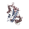 7vuoC  7vvhC  4v0cS S: Starting model for refinement C: citing same article ( |
|---|---|
| Similar structure data |
-Links
 PDBj
PDBj


- Assembly
| Deposited unit | 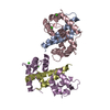
| ||||||||
|---|---|---|---|---|---|---|---|---|---|
| 1 | 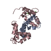
| ||||||||
| 2 | 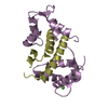
| ||||||||
| Unit cell |
|
-Components
| #1: Protein | Mass: 8122.424 Da / Num. of mol.: 2 / Fragment: C-terminal Domain Source method: isolated from a genetically manipulated source Source: (gene. exp.) Homo sapiens (human) / Gene: KCNQ1, KCNA8, KCNA9, KVLQT1 / Production host:  Escherichia coli (E. coli) / References: UniProt: P51787 Escherichia coli (E. coli) / References: UniProt: P51787#2: Protein | Mass: 16821.531 Da / Num. of mol.: 2 / Mutation: Q135P Source method: isolated from a genetically manipulated source Source: (gene. exp.) Homo sapiens (human) / Gene: CALM1, CALM, CAM, CAM1 / Production host: Escherichia coli (E. coli) / References: UniProt: P0DP23#3: Chemical | ChemComp-CA /   Mass: 40.078 Da / Num. of mol.: 4 / Source method: obtained synthetically / Formula: Ca / Feature type: SUBJECT OF INVESTIGATION Mass: 40.078 Da / Num. of mol.: 4 / Source method: obtained synthetically / Formula: Ca / Feature type: SUBJECT OF INVESTIGATION#4: Water | ChemComp-HOH / | Water Mass: 18.015 Da / Num. of mol.: 2 / Source method: isolated from a natural source / Formula: H2O Mass: 18.015 Da / Num. of mol.: 2 / Source method: isolated from a natural source / Formula: H2OHas ligand of interest | Y | |
|---|
-Experimental details
-Experiment
| Experiment | Method: X-RAY DIFFRACTION / Number of used crystals: 1 |
|---|
- Sample preparation
| Crystal | Density Matthews: 2.21 Å3/Da / Density % sol: 44.31 % |
|---|---|
| Crystal grow | Temperature: 291 K / Method: vapor diffusion, hanging drop / pH: 8 / Details: 25% PEG 1500, 0.1M MMT pH 8.0 |
-Data collection
| Diffraction | Mean temperature: 193.15 K / Serial crystal experiment: N | |||||||||||||||||||||||||||||||||||||||||||||||||||||||||||||||||||||||||||||||||||||||||||||||||||||||||||||||||||||||||||||||||||||||||||||||||||||||||||||||||||||||||||||||||||||||||||||
|---|---|---|---|---|---|---|---|---|---|---|---|---|---|---|---|---|---|---|---|---|---|---|---|---|---|---|---|---|---|---|---|---|---|---|---|---|---|---|---|---|---|---|---|---|---|---|---|---|---|---|---|---|---|---|---|---|---|---|---|---|---|---|---|---|---|---|---|---|---|---|---|---|---|---|---|---|---|---|---|---|---|---|---|---|---|---|---|---|---|---|---|---|---|---|---|---|---|---|---|---|---|---|---|---|---|---|---|---|---|---|---|---|---|---|---|---|---|---|---|---|---|---|---|---|---|---|---|---|---|---|---|---|---|---|---|---|---|---|---|---|---|---|---|---|---|---|---|---|---|---|---|---|---|---|---|---|---|---|---|---|---|---|---|---|---|---|---|---|---|---|---|---|---|---|---|---|---|---|---|---|---|---|---|---|---|---|---|---|---|---|
| Diffraction source | Source: SYNCHROTRON / Site: SSRF / Beamline: BL18U1 / Wavelength: 0.9792 Å | |||||||||||||||||||||||||||||||||||||||||||||||||||||||||||||||||||||||||||||||||||||||||||||||||||||||||||||||||||||||||||||||||||||||||||||||||||||||||||||||||||||||||||||||||||||||||||||
| Detector | Type: DECTRIS PILATUS 6M / Detector: PIXEL / Date: Sep 30, 2018 | |||||||||||||||||||||||||||||||||||||||||||||||||||||||||||||||||||||||||||||||||||||||||||||||||||||||||||||||||||||||||||||||||||||||||||||||||||||||||||||||||||||||||||||||||||||||||||||
| Radiation | Protocol: SINGLE WAVELENGTH / Monochromatic (M) / Laue (L): M / Scattering type: x-ray | |||||||||||||||||||||||||||||||||||||||||||||||||||||||||||||||||||||||||||||||||||||||||||||||||||||||||||||||||||||||||||||||||||||||||||||||||||||||||||||||||||||||||||||||||||||||||||||
| Radiation wavelength | Wavelength: 0.9792 Å / Relative weight: 1 | |||||||||||||||||||||||||||||||||||||||||||||||||||||||||||||||||||||||||||||||||||||||||||||||||||||||||||||||||||||||||||||||||||||||||||||||||||||||||||||||||||||||||||||||||||||||||||||
| Reflection | Resolution: 3.134→50 Å / Num. obs: 7537 / % possible obs: 93.3 % / Redundancy: 4.9 % / Rmerge(I) obs: 0.117 / Rpim(I) all: 0.058 / Rrim(I) all: 0.131 / Χ2: 0.804 / Net I/σ(I): 5.9 | |||||||||||||||||||||||||||||||||||||||||||||||||||||||||||||||||||||||||||||||||||||||||||||||||||||||||||||||||||||||||||||||||||||||||||||||||||||||||||||||||||||||||||||||||||||||||||||
| Reflection shell | Diffraction-ID: 1
|
- Processing
| Software |
| ||||||||||||||||||||||||||||||||||||
|---|---|---|---|---|---|---|---|---|---|---|---|---|---|---|---|---|---|---|---|---|---|---|---|---|---|---|---|---|---|---|---|---|---|---|---|---|---|
| Refinement | Method to determine structure: MOLECULAR REPLACEMENT Starting model: 4V0C Resolution: 3.134→37.793 Å / SU ML: 0.45 / Cross valid method: THROUGHOUT / σ(F): 1.34 / Phase error: 28.91 / Stereochemistry target values: ML
| ||||||||||||||||||||||||||||||||||||
| Solvent computation | Shrinkage radii: 0.9 Å / VDW probe radii: 1.11 Å / Solvent model: FLAT BULK SOLVENT MODEL | ||||||||||||||||||||||||||||||||||||
| Displacement parameters | Biso max: 125.34 Å2 / Biso mean: 57.1726 Å2 / Biso min: 39.48 Å2 | ||||||||||||||||||||||||||||||||||||
| Refinement step | Cycle: final / Resolution: 3.134→37.793 Å
| ||||||||||||||||||||||||||||||||||||
| LS refinement shell | Refine-ID: X-RAY DIFFRACTION / Rfactor Rfree error: 0
|
