 Movie
Movie Controller

[English] 日本語
 Yorodumi
- PDB-7vs9: Crystal structure of P domain from norovirus GI.9 capsid protein ... -
+ Open data

- Basic information
| Entry | Database: PDB / ID: 7vs9 | ||||||
|---|---|---|---|---|---|---|---|
| Title | Crystal structure of P domain from norovirus GI.9 capsid protein in complex with Lewis x antigen. | ||||||
 Components Components | VP1 | ||||||
 Keywords Keywords |  VIRAL PROTEIN / norovirus / p-domain / capsid / histo blood group antigen VIRAL PROTEIN / norovirus / p-domain / capsid / histo blood group antigen | ||||||
| Function / homology | Calicivirus coat protein C-terminal / Calicivirus coat protein C-terminal / Calicivirus coat protein / Calicivirus coat protein / Picornavirus/Calicivirus coat protein / Viral coat protein subunit / Lewis X antigen, alpha anomer / VP1 Function and homology information Function and homology information | ||||||
| Biological species |  Norovirus Hu/GI/Vancouver730/2004/CAN Norovirus Hu/GI/Vancouver730/2004/CAN | ||||||
| Method | X-RAY DIFFRACTION / MOLECULAR REPLACEMENT / Resolution: 2.26 Å | ||||||
 Authors Authors | Murayama, K. / Kato-Murayama, M. / Shirouzu, M. | ||||||
| Funding support |  Japan, 1items Japan, 1items
| ||||||
 Citation Citation | Journal: Febs Open Bio / Year: 2022 Title: Lewis fucose is a key moiety for the recognition of histo-blood group antigens by GI.9 norovirus, as revealed by structural analysis. Authors: Kimura-Someya, T. / Kato-Murayama, M. / Katsura, K. / Sakai, N. / Murayama, K. / Hanada, K. / Shirouzu, M. / Someya, Y. | ||||||
| History |
|
- Structure visualization
| Structure viewer | Molecule: MolmilJmol/JSmol |
|---|
- Downloads & links
-Download
| PDBx/mmCIF format | 7vs9.cif.gz | 147.5 KB | Display | PDBx/mmCIF format |
|---|---|---|---|---|
| PDB format | pdb7vs9.ent.gz | 112 KB | Display | PDB format |
| PDBx/mmJSON format | 7vs9.json.gz | Tree view | PDBx/mmJSON format | |
| Others |  Other downloads Other downloads |
-Validation report
| Arichive directory | https://data.pdbj.org/pub/pdb/validation_reports/vs/7vs9ftp://data.pdbj.org/pub/pdb/validation_reports/vs/7vs9 | HTTPS FTP |
|---|
-Related structure data
| Related structure data | 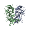 7vp0SC 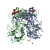 7vs8C S: Starting model for refinement C: citing same article ( |
|---|---|
| Similar structure data |
-Links
 PDBj
PDBj
- Assembly
| Deposited unit | 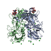
| ||||||||
|---|---|---|---|---|---|---|---|---|---|
| 1 |
| ||||||||
| Unit cell |
|
-Components
| #1: Protein | Mass: 33777.852 Da / Num. of mol.: 2 Source method: isolated from a genetically manipulated source Source: (gene. exp.) Norovirus Hu/GI/Vancouver730/2004/CAN / Details (production host): Cell-free protein synthesis / Production host:  Escherichia coli (E. coli) / References: UniProt: F2XMU3 Escherichia coli (E. coli) / References: UniProt: F2XMU3#2: Polysaccharide | 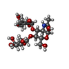  , Oligosaccharide / Class: Antigen / Mass: 529.490 Da / Num. of mol.: 2 , Oligosaccharide / Class: Antigen / Mass: 529.490 Da / Num. of mol.: 2Source method: isolated from a genetically manipulated source Details: oligosaccharide with branches / References: Lewis X antigen, alpha anomer #3: Chemical |   Mass: 24.305 Da / Num. of mol.: 2 / Source method: obtained synthetically / Formula: Mg Mass: 24.305 Da / Num. of mol.: 2 / Source method: obtained synthetically / Formula: Mg#4: Chemical | ChemComp-CL / Chloride  Mass: 35.453 Da / Num. of mol.: 16 / Source method: obtained synthetically / Formula: Cl Mass: 35.453 Da / Num. of mol.: 16 / Source method: obtained synthetically / Formula: Cl#5: Water | ChemComp-HOH / | Water Mass: 18.015 Da / Num. of mol.: 523 / Source method: isolated from a natural source / Formula: H2O Mass: 18.015 Da / Num. of mol.: 523 / Source method: isolated from a natural source / Formula: H2OHas ligand of interest | Y | |
|---|
-Experimental details
-Experiment
| Experiment | Method: X-RAY DIFFRACTION / Number of used crystals: 1 |
|---|
- Sample preparation
| Crystal | Density Matthews: 4.51 Å3/Da / Density % sol: 72.72 % |
|---|---|
| Crystal grow | Temperature: 298 K / Method: vapor diffusion, sitting drop / pH: 9 / Details: magnesium chloride, Bicine, pH 9.0 |
-Data collection
| Diffraction | Mean temperature: 100 K / Serial crystal experiment: N |
|---|---|
| Diffraction source | Source: ROTATING ANODE / Type: RIGAKU FR-E SUPERBRIGHT / Wavelength: 1.5418 Å |
| Detector | Type: RIGAKU RAXIS IV++ / Detector: IMAGE PLATE / Date: Feb 27, 2019 |
| Radiation | Protocol: SINGLE WAVELENGTH / Monochromatic (M) / Laue (L): M / Scattering type: x-ray |
| Radiation wavelength | Wavelength: 1.5418 Å / Relative weight: 1 |
| Reflection | Resolution: 2.26→50 Å / Num. obs: 56256 / % possible obs: 99.6 % / Redundancy: 3.7 % / Rmerge(I) obs: 0.088 / Rrim(I) all: 0.103 / Net I/σ(I): 15.3 |
| Reflection shell | Resolution: 2.26→2.34 Å / Rmerge(I) obs: 0.349 / Num. unique obs: 5539 / Rrim(I) all: 0.411 |
- Processing
| Software |
| ||||||||||||||||||||||||||||||||||||||||||||||||||||||||||||||||||||||||||||||||||||||||||||||||||
|---|---|---|---|---|---|---|---|---|---|---|---|---|---|---|---|---|---|---|---|---|---|---|---|---|---|---|---|---|---|---|---|---|---|---|---|---|---|---|---|---|---|---|---|---|---|---|---|---|---|---|---|---|---|---|---|---|---|---|---|---|---|---|---|---|---|---|---|---|---|---|---|---|---|---|---|---|---|---|---|---|---|---|---|---|---|---|---|---|---|---|---|---|---|---|---|---|---|---|---|
| Refinement | Method to determine structure: MOLECULAR REPLACEMENT Starting model: 7VP0 Resolution: 2.26→35.207 Å / SU ML: 0.21 / Cross valid method: FREE R-VALUE / σ(F): 1.36 / Phase error: 19.87 / Stereochemistry target values: ML
| ||||||||||||||||||||||||||||||||||||||||||||||||||||||||||||||||||||||||||||||||||||||||||||||||||
| Solvent computation | Shrinkage radii: 0.9 Å / VDW probe radii: 1.11 Å / Solvent model: FLAT BULK SOLVENT MODEL | ||||||||||||||||||||||||||||||||||||||||||||||||||||||||||||||||||||||||||||||||||||||||||||||||||
| Refinement step | Cycle: LAST / Resolution: 2.26→35.207 Å
| ||||||||||||||||||||||||||||||||||||||||||||||||||||||||||||||||||||||||||||||||||||||||||||||||||
| Refine LS restraints |
| ||||||||||||||||||||||||||||||||||||||||||||||||||||||||||||||||||||||||||||||||||||||||||||||||||
| LS refinement shell |
|
