 Movie
Movie Controller


+ Open data

- Basic information
| Entry | Database: PDB / ID: 7vs4 | ||||||||||||
|---|---|---|---|---|---|---|---|---|---|---|---|---|---|
| Title | Crystal structure of PacII_M1M2S-DNA(m6A)-SAH complex | ||||||||||||
 Components Components |
| ||||||||||||
 Keywords Keywords | TRANSFERASE/DNA / Type I R-M system PacII methytransferase m4C and m6A modification complex /  BIOSYNTHETIC PROTEIN / TRANSFERASE-DNA complex BIOSYNTHETIC PROTEIN / TRANSFERASE-DNA complex | ||||||||||||
| Function / homology |  Function and homology information Function and homology informationN-methyltransferase activity / site-specific DNA-methyltransferase (adenine-specific) / site-specific DNA-methyltransferase (adenine-specific) activity / DNA bindingSimilarity search - Function | ||||||||||||
| Biological species |  Pseudomonas alcaligenes (bacteria) Pseudomonas alcaligenes (bacteria)synthetic construct (others) | ||||||||||||
| Method | X-RAY DIFFRACTION / SYNCHROTRON / MOLECULAR REPLACEMENT / Resolution: 2.55 Å | ||||||||||||
 Authors Authors | Zhu, J. / Gao, P. | ||||||||||||
| Funding support |  China, 3items China, 3items
| ||||||||||||
 Citation Citation | Journal: Nat Commun / Year: 2022 Title: Molecular insights into DNA recognition and methylation by non-canonical type I restriction-modification systems. Authors: Zhu, J. / Gao, Y. / Wang, Y. / Zhan, Q. / Feng, H. / Luo, X. / Li, P. / Liu, S. / Hou, H. / Gao, P. #1: Journal: Acta Crystallogr., Sect. D: Biol. Crystallogr. / Year: 2012Title: Structural basis underlying complex assembly and conformational transition of the type I R-M system Authors: Yanping, L. / Xiaoxue, Y. | ||||||||||||
| History |
|
- Structure visualization
| Structure viewer | Molecule: MolmilJmol/JSmol |
|---|
- Downloads & links
-Download
| PDBx/mmCIF format | 7vs4.cif.gz | 742.1 KB | Display | PDBx/mmCIF format |
|---|---|---|---|---|
| PDB format | pdb7vs4.ent.gz | 504.4 KB | Display | PDB format |
| PDBx/mmJSON format | 7vs4.json.gz | Tree view | PDBx/mmJSON format | |
| Others |  Other downloads Other downloads |
-Validation report
| Arichive directory | https://data.pdbj.org/pub/pdb/validation_reports/vs/7vs4ftp://data.pdbj.org/pub/pdb/validation_reports/vs/7vs4 | HTTPS FTP |
|---|
-Related structure data
| Related structure data |  7vruSC S: Starting model for refinement C: citing same article ( |
|---|---|
| Similar structure data |
-Links
 PDBj
PDBj


- Assembly
| Deposited unit | 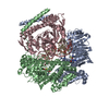
| ||||||||||||
|---|---|---|---|---|---|---|---|---|---|---|---|---|---|
| 1 |
| ||||||||||||
| Unit cell |
|
-Components
-Site-specific DNA-methyltransferase (adenine- ... , 2 types, 2 molecules AB
| #1: Protein | DNA methyltransferase / M1 Mass: 56572.133 Da / Num. of mol.: 1 Source method: isolated from a genetically manipulated source Source: (gene. exp.) Pseudomonas alcaligenes (bacteria) / Gene: A0T30_13480 / Production host: Escherichia coli (E. coli) / Strain (production host): DE3References: UniProt: A0A142ISP4, site-specific DNA-methyltransferase (adenine-specific) |
|---|---|
| #2: Protein | DNA methyltransferase Mass: 57607.355 Da / Num. of mol.: 1 Source method: isolated from a genetically manipulated source Source: (gene. exp.) Pseudomonas alcaligenes (bacteria) / Gene: A0T30_13470 / Production host: Escherichia coli (E. coli) / Strain (production host): DE3References: UniProt: A0A142ISP2, site-specific DNA-methyltransferase (adenine-specific) |
-Protein , 1 types, 1 molecules C
| #3: Protein | Mass: 42852.215 Da / Num. of mol.: 1 Source method: isolated from a genetically manipulated source Source: (gene. exp.) Pseudomonas alcaligenes (bacteria) / Gene: pacIIS / Production host: Escherichia coli (E. coli) / Strain (production host): DE3 |
|---|
-DNA chain , 2 types, 2 molecules HI
| #4: DNA chain | Mass: 7620.953 Da / Num. of mol.: 1 / Source method: obtained synthetically / Source: (synth.) synthetic construct (others) |
|---|---|
| #5: DNA chain | Mass: 7750.004 Da / Num. of mol.: 1 / Source method: obtained synthetically / Source: (synth.) synthetic construct (others) |
-Non-polymers , 2 types, 421 molecules 

| #6: Chemical | S-Adenosyl-L-homocysteine Type: L-peptide linking / Mass: 384.411 Da / Num. of mol.: 2 / Source method: obtained synthetically / Formula: C14H20N6O5S / Feature type: SUBJECT OF INVESTIGATION Type: L-peptide linking / Mass: 384.411 Da / Num. of mol.: 2 / Source method: obtained synthetically / Formula: C14H20N6O5S / Feature type: SUBJECT OF INVESTIGATION#7: Water | ChemComp-HOH / | WaterMass: 18.015 Da / Num. of mol.: 419 / Source method: isolated from a natural source / Formula: H2O |
|---|
-Details
| Has ligand of interest | Y |
|---|
-Experimental details
-Experiment
| Experiment | Method: X-RAY DIFFRACTION / Number of used crystals: 1 |
|---|
- Sample preparation
| Crystal | Density Matthews: 2.73 Å3/Da / Density % sol: 55 % / Description: biconical or anomaly cylindrical object |
|---|---|
| Crystal grow | Temperature: 293 K / Method: vapor diffusion, hanging drop Details: 0.15 M di-ammonium hydrogen phosphate, 25% PEG 3350, 0.2M Li2SO4, 0.1 M Tris pH 8.5, 20% PEG 3350 (another condition) PH range: 6.7-7.2 |
-Data collection
| Diffraction | Mean temperature: 80 K / Serial crystal experiment: N |
|---|---|
| Diffraction source | Source: SYNCHROTRON / Site: SSRF / Beamline: BL18U1 / Wavelength: 0.979 Å |
| Detector | Type: DECTRIS EIGER R 4M / Detector: PIXEL / Date: Oct 3, 2021 |
| Radiation | Protocol: MAD / Monochromatic (M) / Laue (L): M / Scattering type: x-ray |
| Radiation wavelength | Wavelength: 0.979 Å / Relative weight: 1 |
| Reflection | Resolution: 2.55→49.17 Å / Num. obs: 56313 / % possible obs: 98.84 % / Redundancy: 1.6 % / Biso Wilson estimate: 35.96 Å2 / CC1/2: 0.998 / CC star: 1 / Rmerge(I) obs: 0.02371 / Rpim(I) all: 0.02317 / Rrim(I) all: 0.03277 / Net I/σ(I): 18.54 |
| Reflection shell | Resolution: 2.55→2.641 Å / Rmerge(I) obs: 0.319 / Mean I/σ(I) obs: 2.71 / Num. unique obs: 5361 / CC1/2: 1 / CC star: 1 / Rpim(I) all: 0.319 / Rrim(I) all: 0.4352 |
- Processing
| Software |
| |||||||||||||||||||||||||||||||||||||||||||||||||||||||||||||||||||||||||||||||||||||||||||||||||||||||||||||||||||||||||||||||||||||||||||||||||||
|---|---|---|---|---|---|---|---|---|---|---|---|---|---|---|---|---|---|---|---|---|---|---|---|---|---|---|---|---|---|---|---|---|---|---|---|---|---|---|---|---|---|---|---|---|---|---|---|---|---|---|---|---|---|---|---|---|---|---|---|---|---|---|---|---|---|---|---|---|---|---|---|---|---|---|---|---|---|---|---|---|---|---|---|---|---|---|---|---|---|---|---|---|---|---|---|---|---|---|---|---|---|---|---|---|---|---|---|---|---|---|---|---|---|---|---|---|---|---|---|---|---|---|---|---|---|---|---|---|---|---|---|---|---|---|---|---|---|---|---|---|---|---|---|---|---|---|---|---|
| Refinement | Method to determine structure: MOLECULAR REPLACEMENT Starting model: 7VRU Resolution: 2.55→49.17 Å / SU ML: 0.2978 / Cross valid method: FREE R-VALUE / σ(F): 1.34 / Phase error: 23.3725 Stereochemistry target values: GeoStd + Monomer Library + CDL v1.2
| |||||||||||||||||||||||||||||||||||||||||||||||||||||||||||||||||||||||||||||||||||||||||||||||||||||||||||||||||||||||||||||||||||||||||||||||||||
| Solvent computation | Shrinkage radii: 0.9 Å / VDW probe radii: 1.11 Å / Solvent model: FLAT BULK SOLVENT MODEL | |||||||||||||||||||||||||||||||||||||||||||||||||||||||||||||||||||||||||||||||||||||||||||||||||||||||||||||||||||||||||||||||||||||||||||||||||||
| Displacement parameters | Biso mean: 40.24 Å2 | |||||||||||||||||||||||||||||||||||||||||||||||||||||||||||||||||||||||||||||||||||||||||||||||||||||||||||||||||||||||||||||||||||||||||||||||||||
| Refinement step | Cycle: LAST / Resolution: 2.55→49.17 Å
| |||||||||||||||||||||||||||||||||||||||||||||||||||||||||||||||||||||||||||||||||||||||||||||||||||||||||||||||||||||||||||||||||||||||||||||||||||
| Refine LS restraints |
| |||||||||||||||||||||||||||||||||||||||||||||||||||||||||||||||||||||||||||||||||||||||||||||||||||||||||||||||||||||||||||||||||||||||||||||||||||
| LS refinement shell |
| |||||||||||||||||||||||||||||||||||||||||||||||||||||||||||||||||||||||||||||||||||||||||||||||||||||||||||||||||||||||||||||||||||||||||||||||||||
| Refinement TLS params. | Method: refined / Origin x: 9.85819719049 Å / Origin y: 69.3043024957 Å / Origin z: 90.020295974 Å
| |||||||||||||||||||||||||||||||||||||||||||||||||||||||||||||||||||||||||||||||||||||||||||||||||||||||||||||||||||||||||||||||||||||||||||||||||||
| Refinement TLS group | Selection details: all |
