 Movie
Movie Controller

[English] 日本語
 Yorodumi
- PDB-7vrd: Crystal structure of Enolase1 from Candida albicans complexed wit... -
+ Open data

- Basic information
| Entry | Database: PDB / ID: 7vrd | ||||||
|---|---|---|---|---|---|---|---|
| Title | Crystal structure of Enolase1 from Candida albicans complexed with 2'-phosphoglyceric acid sodium | ||||||
 Components Components | Enolase 1 Alpha-enolase Alpha-enolase | ||||||
 Keywords Keywords | METAL BINDING PROTEIN | ||||||
| Function / homology |  Function and homology information Function and homology informationfungal-type cell wall organization or biogenesis / high molecular weight kininogen binding / filamentous growth of a population of unicellular organisms in response to biotic stimulus / yeast-form cell wall / : / fungal biofilm matrix / hyphal cell wall / protein-glutamine gamma-glutamyltransferase activity / phosphopyruvate hydratase / phosphopyruvate hydratase complex ...fungal-type cell wall organization or biogenesis / high molecular weight kininogen binding / filamentous growth of a population of unicellular organisms in response to biotic stimulus / yeast-form cell wall / : / fungal biofilm matrix / hyphal cell wall / protein-glutamine gamma-glutamyltransferase activity / phosphopyruvate hydratase / phosphopyruvate hydratase complex / phosphopyruvate hydratase activity / fungal-type cell wall / symbiont entry into host / filamentous growth / fibrinolysis / gluconeogenesis / glycolytic process / extracellular vesicle / external side of plasma membrane / magnesium ion binding / cell surface / extracellular region / membrane / nucleus / plasma membrane / cytosolSimilarity search - Function | ||||||
| Biological species |  Candida albicans SC5314 (yeast) Candida albicans SC5314 (yeast) | ||||||
| Method | X-RAY DIFFRACTION / SYNCHROTRON / MOLECULAR REPLACEMENT / Resolution: 1.7 Å | ||||||
 Authors Authors | Zhang, M. / Zhang, X. | ||||||
| Funding support |  China, 1items China, 1items
| ||||||
 Citation Citation | Journal: Microbiol Spectr / Year: 2022 Title: Baicalein Acts against Candida albicans by Targeting Eno1 and Inhibiting Glycolysis. Authors: Li, L. / Lu, H. / Zhang, X. / Whiteway, M. / Wu, H. / Tan, S. / Zang, J. / Tian, S. / Zhen, C. / Meng, X. / Li, W. / Zhang, D. / Zhang, M. / Jiang, Y. | ||||||
| History |
|
- Structure visualization
| Structure viewer | Molecule: MolmilJmol/JSmol |
|---|
- Downloads & links
-Download
| PDBx/mmCIF format | 7vrd.cif.gz | 357.2 KB | Display | PDBx/mmCIF format |
|---|---|---|---|---|
| PDB format | pdb7vrd.ent.gz | 288.3 KB | Display | PDB format |
| PDBx/mmJSON format | 7vrd.json.gz | Tree view | PDBx/mmJSON format | |
| Others |  Other downloads Other downloads |
-Validation report
| Arichive directory | https://data.pdbj.org/pub/pdb/validation_reports/vr/7vrdftp://data.pdbj.org/pub/pdb/validation_reports/vr/7vrd | HTTPS FTP |
|---|
-Related structure data
| Related structure data | 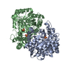 7v67C  2al2S S: Starting model for refinement C: citing same article ( |
|---|---|
| Similar structure data |
-Links
 PDBj
PDBj
- Assembly
| Deposited unit | 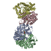
| ||||||||
|---|---|---|---|---|---|---|---|---|---|
| 1 | 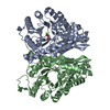
| ||||||||
| 2 | 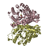
| ||||||||
| Unit cell |
|
-Components
| #1: Protein | Alpha-enolase / 2-phospho-D-glycerate hydro-lyase / 2-phosphoglycerate dehydratase Mass: 47156.156 Da / Num. of mol.: 4 Source method: isolated from a genetically manipulated source Source: (gene. exp.) Candida albicans SC5314 (yeast) / Strain: SC5314 / Gene: ENO1 / Production host:  Escherichia coli (E. coli) / References: UniProt: P30575, phosphopyruvate hydratase Escherichia coli (E. coli) / References: UniProt: P30575, phosphopyruvate hydratase#2: Chemical | ChemComp-MG /   Mass: 24.305 Da / Num. of mol.: 6 / Source method: obtained synthetically / Formula: Mg Mass: 24.305 Da / Num. of mol.: 6 / Source method: obtained synthetically / Formula: Mg#3: Chemical | 2-Phosphoglyceric acid  Mass: 186.057 Da / Num. of mol.: 2 / Source method: obtained synthetically / Formula: C3H7O7P / Feature type: SUBJECT OF INVESTIGATION Mass: 186.057 Da / Num. of mol.: 2 / Source method: obtained synthetically / Formula: C3H7O7P / Feature type: SUBJECT OF INVESTIGATION#4: Water | ChemComp-HOH / | Water Mass: 18.015 Da / Num. of mol.: 1205 / Source method: isolated from a natural source / Formula: H2O Mass: 18.015 Da / Num. of mol.: 1205 / Source method: isolated from a natural source / Formula: H2OHas ligand of interest | Y | |
|---|
-Experimental details
-Experiment
| Experiment | Method: X-RAY DIFFRACTION / Number of used crystals: 1 |
|---|
- Sample preparation
| Crystal | Density Matthews: 2.39 Å3/Da / Density % sol: 48.5 % |
|---|---|
| Crystal grow | Temperature: 293 K / Method: vapor diffusion, sitting drop Details: 0.2 M Magnesium acetate tetrahydrate, 0.1 M Sodium cacodylate trihydrate pH 6.5, 20% w/v Polyethlene glycol 8,000 |
-Data collection
| Diffraction | Mean temperature: 100 K / Serial crystal experiment: N | |||||||||||||||||||||||||||||||||||||||||||||||||||||||||||||||||||||||||||||||||||||||||||||||||||
|---|---|---|---|---|---|---|---|---|---|---|---|---|---|---|---|---|---|---|---|---|---|---|---|---|---|---|---|---|---|---|---|---|---|---|---|---|---|---|---|---|---|---|---|---|---|---|---|---|---|---|---|---|---|---|---|---|---|---|---|---|---|---|---|---|---|---|---|---|---|---|---|---|---|---|---|---|---|---|---|---|---|---|---|---|---|---|---|---|---|---|---|---|---|---|---|---|---|---|---|---|
| Diffraction source | Source: SYNCHROTRON / Site: SSRF / Beamline: BL17U1 / Wavelength: 0.979 Å | |||||||||||||||||||||||||||||||||||||||||||||||||||||||||||||||||||||||||||||||||||||||||||||||||||
| Detector | Type: ADSC QUANTUM 315r / Detector: CCD / Date: Jan 16, 2021 | |||||||||||||||||||||||||||||||||||||||||||||||||||||||||||||||||||||||||||||||||||||||||||||||||||
| Radiation | Protocol: SINGLE WAVELENGTH / Monochromatic (M) / Laue (L): M / Scattering type: x-ray | |||||||||||||||||||||||||||||||||||||||||||||||||||||||||||||||||||||||||||||||||||||||||||||||||||
| Radiation wavelength | Wavelength: 0.979 Å / Relative weight: 1 | |||||||||||||||||||||||||||||||||||||||||||||||||||||||||||||||||||||||||||||||||||||||||||||||||||
| Reflection | Resolution: 1.7→50 Å / Num. obs: 185469 / % possible obs: 99.6 % / Redundancy: 6.7 % / Rmerge(I) obs: 0.069 / Rpim(I) all: 0.029 / Rrim(I) all: 0.075 / Χ2: 0.738 / Net I/σ(I): 7.7 / Num. measured all: 1237061 | |||||||||||||||||||||||||||||||||||||||||||||||||||||||||||||||||||||||||||||||||||||||||||||||||||
| Reflection shell | Diffraction-ID: 1
|
- Processing
| Software |
| ||||||||||||||||||||||||||||||||||||||||||||||||||||||||||||
|---|---|---|---|---|---|---|---|---|---|---|---|---|---|---|---|---|---|---|---|---|---|---|---|---|---|---|---|---|---|---|---|---|---|---|---|---|---|---|---|---|---|---|---|---|---|---|---|---|---|---|---|---|---|---|---|---|---|---|---|---|---|
| Refinement | Method to determine structure: MOLECULAR REPLACEMENT Starting model: 2AL2 Resolution: 1.7→38.06 Å / Cor.coef. Fo:Fc: 0.966 / Cor.coef. Fo:Fc free: 0.958 / SU B: 1.781 / SU ML: 0.06 / Cross valid method: THROUGHOUT / σ(F): 0 / ESU R: 0.104 / ESU R Free: 0.098 / Stereochemistry target values: MAXIMUM LIKELIHOOD Details: HYDROGENS HAVE BEEN ADDED IN THE RIDING POSITIONS U VALUES : REFINED INDIVIDUALLY
| ||||||||||||||||||||||||||||||||||||||||||||||||||||||||||||
| Solvent computation | Ion probe radii: 0.8 Å / Shrinkage radii: 0.8 Å / VDW probe radii: 1.2 Å / Solvent model: MASK | ||||||||||||||||||||||||||||||||||||||||||||||||||||||||||||
| Displacement parameters | Biso max: 58.37 Å2 / Biso mean: 18.906 Å2 / Biso min: 7.74 Å2
| ||||||||||||||||||||||||||||||||||||||||||||||||||||||||||||
| Refinement step | Cycle: final / Resolution: 1.7→38.06 Å
| ||||||||||||||||||||||||||||||||||||||||||||||||||||||||||||
| Refine LS restraints |
| ||||||||||||||||||||||||||||||||||||||||||||||||||||||||||||
| LS refinement shell | Resolution: 1.701→1.746 Å / Rfactor Rfree error: 0 / Total num. of bins used: 20
|
