 Movie
Movie Controller


+ Open data

- Basic information
| Entry | Database: PDB / ID: 7vp1 | |||||||||
|---|---|---|---|---|---|---|---|---|---|---|
| Title | Structure of a transcription factor and DNA complex | |||||||||
 Components Components |
| |||||||||
 Keywords Keywords |  DNA BINDING PROTEIN / Complex / Transcription factor / TRANSCRIPTION DNA BINDING PROTEIN / Complex / Transcription factor / TRANSCRIPTION | |||||||||
| Function / homology |  Function and homology information Function and homology informationleaf senescence / leaf development / DNA-binding transcription factor activity / regulation of DNA-templated transcription / DNA binding / nucleusSimilarity search - Function | |||||||||
| Biological species |  Arabidopsis thaliana (thale cress) Arabidopsis thaliana (thale cress)synthetic construct (others) | |||||||||
| Method | X-RAY DIFFRACTION / SYNCHROTRON / SAD / Resolution: 2.902 Å | |||||||||
 Authors Authors | Zhang, Y. / Xu, Y.P. / Wang, B. / Su, X.D. | |||||||||
| Funding support |  China, 2items China, 2items
| |||||||||
 Citation Citation | Journal: To Be Published Title: Structural basis for DNA recognition by TCP transcription factors Authors: Zhang, Y. / Xu, Y.P. / Wang, B. / Su, X.D. | |||||||||
| History |
|
- Structure visualization
| Structure viewer | Molecule: MolmilJmol/JSmol |
|---|
- Downloads & links
-Download
| PDBx/mmCIF format | 7vp1.cif.gz | 85.9 KB | Display | PDBx/mmCIF format |
|---|---|---|---|---|
| PDB format | pdb7vp1.ent.gz | 64.8 KB | Display | PDB format |
| PDBx/mmJSON format | 7vp1.json.gz | Tree view | PDBx/mmJSON format | |
| Others |  Other downloads Other downloads |
-Validation report
| Arichive directory | https://data.pdbj.org/pub/pdb/validation_reports/vp/7vp1ftp://data.pdbj.org/pub/pdb/validation_reports/vp/7vp1 | HTTPS FTP |
|---|
-Related structure data
| Related structure data | 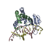 7vp2C 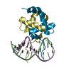 7vp3C 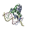 7vp4C 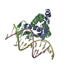 7vp5C  7vp6C 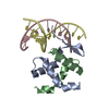 7vp7C C: citing same article ( |
|---|---|
| Similar structure data |
-Links
 PDBj
PDBj


- Assembly
| Deposited unit | 
| ||||||||
|---|---|---|---|---|---|---|---|---|---|
| 1 |
| ||||||||
| Unit cell |
|
-Components
| #1: Protein | Mass: 12045.242 Da / Num. of mol.: 2 Source method: isolated from a genetically manipulated source Source: (gene. exp.) Arabidopsis thaliana (thale cress) / Gene: TCP10, At2g31070, T16B12.12 / Production host:  Escherichia coli BL21(DE3) (bacteria) / References: UniProt: O82277 Escherichia coli BL21(DE3) (bacteria) / References: UniProt: O82277#2: DNA chain | | Mass: 3614.354 Da / Num. of mol.: 1 / Source method: obtained synthetically / Source: (synth.) synthetic construct (others) #3: DNA chain | | Mass: 3712.430 Da / Num. of mol.: 1 / Source method: obtained synthetically / Source: (synth.) synthetic construct (others) Has ligand of interest | N | |
|---|
-Experimental details
-Experiment
| Experiment | Method: X-RAY DIFFRACTION / Number of used crystals: 1 |
|---|
- Sample preparation
| Crystal | Density Matthews: 2.27 Å3/Da / Density % sol: 45.89 % |
|---|---|
| Crystal grow | Temperature: 291 K / Method: vapor diffusion, sitting drop Details: 0.2 M Potassium sodium tartrate tetrahydrate, 20% w/v Polyethylene glycol 3350 |
-Data collection
| Diffraction | Mean temperature: 100 K / Serial crystal experiment: N |
|---|---|
| Diffraction source | Source: SYNCHROTRON / Site: SSRF / Beamline: BL17U1 / Wavelength: 0.9792 Å |
| Detector | Type: DECTRIS EIGER X 16M / Detector: PIXEL / Date: Dec 9, 2018 |
| Radiation | Protocol: SINGLE WAVELENGTH / Monochromatic (M) / Laue (L): M / Scattering type: x-ray |
| Radiation wavelength | Wavelength: 0.9792 Å / Relative weight: 1 |
| Reflection | Resolution: 2.902→29.21 Å / Num. obs: 6982 / % possible obs: 99.52 % / Redundancy: 20 % / CC1/2: 0.999 / Rmerge(I) obs: 0.1173 / Rpim(I) all: 0.01942 / Rrim(I) all: 0.1189 / Net I/σ(I): 38.48 |
| Reflection shell | Resolution: 2.902→3.006 Å / Rmerge(I) obs: 0.6148 / Num. unique obs: 653 / CC1/2: 0.975 / Rpim(I) all: 0.09861 / Rrim(I) all: 0.6228 |
- Processing
| Software |
| ||||||||||||||||||||||||||||||||||||||||||||||||||||||||||||
|---|---|---|---|---|---|---|---|---|---|---|---|---|---|---|---|---|---|---|---|---|---|---|---|---|---|---|---|---|---|---|---|---|---|---|---|---|---|---|---|---|---|---|---|---|---|---|---|---|---|---|---|---|---|---|---|---|---|---|---|---|---|
| Refinement | Method to determine structure: SAD / Resolution: 2.902→29.21 Å / SU ML: 0.26 / Cross valid method: THROUGHOUT / Phase error: 20.78 / Stereochemistry target values: ML
| ||||||||||||||||||||||||||||||||||||||||||||||||||||||||||||
| Solvent computation | Shrinkage radii: 0.9 Å / VDW probe radii: 1.11 Å / Solvent model: FLAT BULK SOLVENT MODEL | ||||||||||||||||||||||||||||||||||||||||||||||||||||||||||||
| Displacement parameters | Biso max: 112.5 Å2 / Biso mean: 49.7307 Å2 / Biso min: 16.88 Å2 | ||||||||||||||||||||||||||||||||||||||||||||||||||||||||||||
| Refinement step | Cycle: final / Resolution: 2.902→29.21 Å
| ||||||||||||||||||||||||||||||||||||||||||||||||||||||||||||
| Refine LS restraints |
| ||||||||||||||||||||||||||||||||||||||||||||||||||||||||||||
| LS refinement shell | Refine-ID: X-RAY DIFFRACTION / Rfactor Rfree error: 0
| ||||||||||||||||||||||||||||||||||||||||||||||||||||||||||||
| Refinement TLS params. | Method: refined / Origin x: 0.6027 Å / Origin y: 23.5675 Å / Origin z: 4.5119 Å
| ||||||||||||||||||||||||||||||||||||||||||||||||||||||||||||
| Refinement TLS group |
|
