 Movie
Movie Controller


+ Open data

- Basic information
| Entry | Database: PDB / ID: 7vmf | |||||||||
|---|---|---|---|---|---|---|---|---|---|---|
| Title | Crystal structure of Arabidopsis thaliana HDT2 | |||||||||
 Components Components | Histone deacetylase HDT2 | |||||||||
 Keywords Keywords |  CHAPERONE / HDT / HD-tuin / Nucleoplasmin / Histone chaperone / HD2B / HDT2 CHAPERONE / HDT / HD-tuin / Nucleoplasmin / Histone chaperone / HD2B / HDT2 | |||||||||
| Function / homology |  Function and homology information Function and homology informationpolarity specification of adaxial/abaxial axis / seed dormancy process / DNA-mediated transformation / plant-type cell wall / root development / plant-type vacuole / histone deacetylase activity / plastid / chromatin organization / negative regulation of DNA-templated transcription ...polarity specification of adaxial/abaxial axis / seed dormancy process / DNA-mediated transformation / plant-type cell wall / root development / plant-type vacuole / histone deacetylase activity / plastid / chromatin organization / negative regulation of DNA-templated transcription / nucleolus / mitochondrion / nucleusSimilarity search - Function | |||||||||
| Biological species |  Arabidopsis thaliana (thale cress) Arabidopsis thaliana (thale cress) | |||||||||
| Method | X-RAY DIFFRACTION / SYNCHROTRON / MOLECULAR REPLACEMENT / Resolution: 1.32 Å | |||||||||
 Authors Authors | Kumar, A. / Bobde, R.C. / Vasudevan, D. | |||||||||
| Funding support |  India, 2items India, 2items
| |||||||||
 Citation Citation | Journal: Plant Cell / Year: 2022 Title: Plant-specific HDT family histone deacetylases are nucleoplasmins. Authors: Bobde, R.C. / Kumar, A. / Vasudevan, D. | |||||||||
| History |
|
- Structure visualization
| Structure viewer | Molecule: MolmilJmol/JSmol |
|---|
- Downloads & links
-Download
| PDBx/mmCIF format | 7vmf.cif.gz | 203 KB | Display | PDBx/mmCIF format |
|---|---|---|---|---|
| PDB format | pdb7vmf.ent.gz | 162.8 KB | Display | PDB format |
| PDBx/mmJSON format | 7vmf.json.gz | Tree view | PDBx/mmJSON format | |
| Others |  Other downloads Other downloads |
-Validation report
| Arichive directory | https://data.pdbj.org/pub/pdb/validation_reports/vm/7vmfftp://data.pdbj.org/pub/pdb/validation_reports/vm/7vmf | HTTPS FTP |
|---|
-Related structure data
| Related structure data | 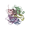 7vmhC 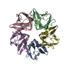 7vmiC 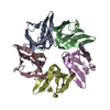 7vrrC  6j2zS S: Starting model for refinement C: citing same article ( |
|---|---|
| Similar structure data |
-Links
 PDBj
PDBj
- Assembly
| Deposited unit | 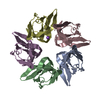
| ||||||||
|---|---|---|---|---|---|---|---|---|---|
| 1 |
| ||||||||
| Unit cell |
|
-Components
| #1: Protein | Mass: 10365.731 Da / Num. of mol.: 5 Source method: isolated from a genetically manipulated source Source: (gene. exp.) Arabidopsis thaliana (thale cress) / Gene: HDT2, HD2, HD2B, HDA4, At5g22650, MDJ22.7 / Plasmid: pET22b / Production host:  Escherichia coli BL21(DE3) (bacteria) / References: UniProt: Q56WH4 Escherichia coli BL21(DE3) (bacteria) / References: UniProt: Q56WH4#2: Chemical | ChemComp-NA / |   Mass: 22.990 Da / Num. of mol.: 1 / Source method: obtained synthetically / Formula: Na Mass: 22.990 Da / Num. of mol.: 1 / Source method: obtained synthetically / Formula: Na#3: Water | ChemComp-HOH / | Water Mass: 18.015 Da / Num. of mol.: 330 / Source method: isolated from a natural source / Formula: H2O Mass: 18.015 Da / Num. of mol.: 330 / Source method: isolated from a natural source / Formula: H2OHas ligand of interest | N | |
|---|
-Experimental details
-Experiment
| Experiment | Method: X-RAY DIFFRACTION / Number of used crystals: 1 |
|---|
- Sample preparation
| Crystal | Density Matthews: 2.44 Å3/Da / Density % sol: 49.6 % |
|---|---|
| Crystal grow | Temperature: 291 K / Method: vapor diffusion, sitting drop / pH: 8 Details: 0.2M ammonium phosphate dibasic pH-8.0, 20%W/V PEG 3350 |
-Data collection
| Diffraction | Mean temperature: 103 K / Serial crystal experiment: N |
|---|---|
| Diffraction source | Source: SYNCHROTRON / Site: ESRF  / Beamline: ID29 / Wavelength: 1.07227 Å / Beamline: ID29 / Wavelength: 1.07227 Å |
| Detector | Type: DECTRIS PILATUS 6M / Detector: PIXEL / Date: Jul 1, 2017 |
| Radiation | Protocol: SINGLE WAVELENGTH / Monochromatic (M) / Laue (L): M / Scattering type: x-ray |
| Radiation wavelength | Wavelength: 1.07227 Å / Relative weight: 1 |
| Reflection | Resolution: 1.32→49.07 Å / Num. obs: 111734 / % possible obs: 95 % / Redundancy: 3.6 % / CC1/2: 0.999 / Net I/σ(I): 13.9 |
| Reflection shell | Resolution: 1.32→1.34 Å / Redundancy: 2.4 % / Mean I/σ(I) obs: 2.1 / Num. unique obs: 4443 / CC1/2: 0.853 / % possible all: 77.2 |
- Processing
| Software |
| |||||||||||||||||||||||||||||||||||||||||||||||||||||||||||||||||||||||||||||||||||||||||||||||||||||||||
|---|---|---|---|---|---|---|---|---|---|---|---|---|---|---|---|---|---|---|---|---|---|---|---|---|---|---|---|---|---|---|---|---|---|---|---|---|---|---|---|---|---|---|---|---|---|---|---|---|---|---|---|---|---|---|---|---|---|---|---|---|---|---|---|---|---|---|---|---|---|---|---|---|---|---|---|---|---|---|---|---|---|---|---|---|---|---|---|---|---|---|---|---|---|---|---|---|---|---|---|---|---|---|---|---|---|---|
| Refinement | Method to determine structure: MOLECULAR REPLACEMENT Starting model: 6J2Z Resolution: 1.32→46.77 Å / Cor.coef. Fo:Fc: 0.972 / Cor.coef. Fo:Fc free: 0.968 / SU B: 1.786 / SU ML: 0.033 / Cross valid method: THROUGHOUT / ESU R: 0.052 / ESU R Free: 0.05 / Stereochemistry target values: MAXIMUM LIKELIHOOD Details: HYDROGENS HAVE BEEN ADDED IN THE RIDING POSITIONS U VALUES : REFINED INDIVIDUALLY
| |||||||||||||||||||||||||||||||||||||||||||||||||||||||||||||||||||||||||||||||||||||||||||||||||||||||||
| Solvent computation | Ion probe radii: 1.1 Å / Shrinkage radii: 1.1 Å / VDW probe radii: 1.4 Å / Solvent model: MASK | |||||||||||||||||||||||||||||||||||||||||||||||||||||||||||||||||||||||||||||||||||||||||||||||||||||||||
| Displacement parameters | Biso mean: 22.148 Å2
| |||||||||||||||||||||||||||||||||||||||||||||||||||||||||||||||||||||||||||||||||||||||||||||||||||||||||
| Refinement step | Cycle: LAST / Resolution: 1.32→46.77 Å
| |||||||||||||||||||||||||||||||||||||||||||||||||||||||||||||||||||||||||||||||||||||||||||||||||||||||||
| Refine LS restraints |
| |||||||||||||||||||||||||||||||||||||||||||||||||||||||||||||||||||||||||||||||||||||||||||||||||||||||||
| LS refinement shell | Resolution: 1.32→1.354 Å / Total num. of bins used: 20
|
